Home > Press > Advanced imaging for bone research and materials science
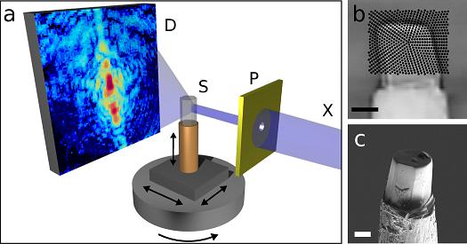 |
| Schematic of the new nano-CT method. The sample is scanned with an X-ray beam while the detector records a diffraction pattern for every beam position. The sample is then turned around its axis and scanned again, until a complete set of data is gathered for every angle. A high-resolution three-dimensional image of the sample is then computed from the hundreds of thousands of diffraction patterns by means of specially developed image reconstruction algorithms. |
Abstract:
High-resolution method for computed nano-tomography developed
Advanced imaging for bone research and materials science
Germany | Posted on October 12th, 2010A novel nano-tomography method developed by a team of researchers from the Technische Universit�t M�nchen (TUM), the Paul Scherrer Institute (PSI) and the ETH Zurich opens the door to computed tomography examinations of minute structures at nanometer resolutions. The new method makes possible, for example, three-dimensional internal imaging of fragile bone structures. The first nano-CT images generated with this procedure will be published in the renowned journal Nature on September 23, 2010. This new technique will facilitate advances in both life sciences and materials sciences.
Osteoporosis, a medical condition in which bones become brittle and fragile from a loss of density, is among the most common diseases in aging bones: In Germany around a quarter of the population aged over 50 is affected. Patients' bone material shrinks rapidly, leading to a significantly increased risk of fracture. In clinical research to date, osteoporosis is diagnosed almost exclusively by establishing an overall reduction in bone density. This approach, however, gives little information about the associated, and equally important, local structure and bone density changes. Franz Pfeiffer, TUM professor for Biomedical Physics and head of the research team, has resolved the dilemma: "With our newly developed nano-CT method it is now possible to visualize the bone structure and density changes at high resolutions and in 3D. This enables us to do research on structural changes related to osteoporosis on a nanoscale and thus develop better therapeutic approaches."
During development, Pfeiffer's team built on X-ray computed tomography (CT). The principle is well established - CT scanners are used every day in hospitals and medical practices for the diagnostic screening of the human body. In the process the human body is X-rayed while a detector records from different angles how much radiation is being absorbed. In principle it is nothing more than taking multiple X-ray pictures from various directions. A number of such pictures are then used to generate digital 3D images of the body's interior using image processing.
The newly developed method measures not only the overall beam intensity absorbed by the object under examination at each angle, but also those parts of the X-ray beam that are deflected in different directions - "diffracted" in the language of physics. Such a diffraction pattern is generated for every point in the sample. This supplies additional information about the exact nanostructure, as X-ray radiation is particularly sensitive to the tiniest of structural changes. "Because we have to take and process so many individual pictures with extreme precision, it was particularly important during the implementation of the method to use high-brilliance X-ray radiation and fast, low-noise pixel detectors - both available at the Swiss Light Source (SLS)," says Oliver Bunk, who was responsible for the requisite experimental setup at the PSI synchrotron facilities in Switzerland.
The diffraction patterns are then processed using an algorithm developed by the team. TUM researcher Martin Dierolf, lead author of the Nature article, explains: "We developed an image reconstruction algorithm that generates a high-resolution, three-dimensional image of the sample using over one hundred thousand diffraction patterns. This algorithm takes into account not only classical X-ray absorption, but also the significantly more sensitive phase shift of the X-rays." A showcase example of the new technique was the examination of a 25-micrometer, superfine bone specimen of a laboratory mouse - with surprisingly exact results. The so-called phase contrast CT pictures show even smallest variations in the specimen's bone density with extremely high precision: Cross-sections of cavities where bone cells reside and their roughly 100 nanometer-fine interconnection network are clearly visible.
"Although the new nano-CT procedure does not achieve the spatial resolution currently available in electron microscopy, it can - because of the high penetration of X-rays - generate three-dimensional tomography images of bone samples," comments Roger Wepf, director of the Electron Microscopy Center of the ETH Zurich (EMEZ). "Furthermore, the new nano-CT procedure stands out with its high precision bone density measurement capacity, which is particularly important in bone research." This method will open the door to more precise studies on the early phase of osteoporosis, in particular, and evaluation of the therapeutic outcomes of various treatments in clinical studies.
The new technique is also very interesting for non-medical applications: Further fields of application include the development of new materials in materials science or in the characterization of semiconductor components. Ultimately, the nano-CT procedure may also be transferred to novel, laser-based X-ray sources, such as the ones currently under development at the Cluster of Excellence "Munich-Centre for Advanced Photonics" (MAP) and at the recently approved large-scale research project "Centre for Advanced Laser Applications" (CALA) on the TUM-Campus Garching near Munich.
Original publication:
Martin Dierolf, Andreas Menzel, Pierre Thibault, Philipp Schneider, Cameron M. Kewish, Roger Wepf, Oliver Bunk, Franz Pfeiffer: "Ptychographic X-Ray Computed Tomography at the Nano-Scale". Nature, 467, 436-439, 23. September 2010 - DOI: 10.1038/nature09419
####
For more information, please click here
Contacts:
Prof. Franz Pfeiffer
Chair of Biomedical Physics Technische Universit�t M�nchen
James-Franck-Stra�e 1 85748 Garching, Germany
Tel.: +49 89 289 12551
Fax: +49 89 289 12548
Dr. Oliver Bunk
Laboratory for Macromolecules and Bioimaging
Paul Scherrer Institute
3232 Villigen PSI, Switzerland
Tel.: +41 56 310 3077
Dr. Roger Albert Wepf
EMEZ � Electron Microscopy ETH Zurich
ETH Zurich
Wolfgang-Pauli-Str. 16 8093 Zurich, Switzerland
Tel: +41 44 633 45 58
Copyright © Technische Universitaet Muenchen
If you have a comment, please Contact us.Issuers of news releases, not 7th Wave, Inc. or Nanotechnology Now, are solely responsible for the accuracy of the content.
Bookmark:


| Related News Press |
News and information
![]() Quantum computer improves AI predictions April 17th, 2026
Quantum computer improves AI predictions April 17th, 2026
![]() Flexible sensor gains sensitivity under pressure April 17th, 2026
Flexible sensor gains sensitivity under pressure April 17th, 2026
![]() A reusable chip for particulate matter sensing April 17th, 2026
A reusable chip for particulate matter sensing April 17th, 2026
![]() Detecting vibrational quantum beating in the predissociation dynamics of SF6 using time-resolved photoelectron spectroscopy April 17th, 2026
Detecting vibrational quantum beating in the predissociation dynamics of SF6 using time-resolved photoelectron spectroscopy April 17th, 2026
Physics
![]() UC Irvine physicists discover method to reverse �quantum scrambling� : The work addresses the problem of information loss in quantum computing system April 17th, 2026
UC Irvine physicists discover method to reverse �quantum scrambling� : The work addresses the problem of information loss in quantum computing system April 17th, 2026
![]() Quantum computers simulate fundamental physics: shedding light on the building blocks of nature June 6th, 2025
Quantum computers simulate fundamental physics: shedding light on the building blocks of nature June 6th, 2025
Possible Futures
![]() A fundamentally new therapeutic approach to cystic fibrosis: Nanobody repairs cellular defect April 17th, 2026
A fundamentally new therapeutic approach to cystic fibrosis: Nanobody repairs cellular defect April 17th, 2026
![]() UC Irvine physicists discover method to reverse �quantum scrambling� : The work addresses the problem of information loss in quantum computing system April 17th, 2026
UC Irvine physicists discover method to reverse �quantum scrambling� : The work addresses the problem of information loss in quantum computing system April 17th, 2026
Academic/Education
![]() Rice University launches Rice Synthetic Biology Institute to improve lives January 12th, 2024
Rice University launches Rice Synthetic Biology Institute to improve lives January 12th, 2024
![]() Multi-institution, $4.6 million NSF grant to fund nanotechnology training September 9th, 2022
Multi-institution, $4.6 million NSF grant to fund nanotechnology training September 9th, 2022
Nanomedicine
![]() A fundamentally new therapeutic approach to cystic fibrosis: Nanobody repairs cellular defect April 17th, 2026
A fundamentally new therapeutic approach to cystic fibrosis: Nanobody repairs cellular defect April 17th, 2026
![]() New molecular technology targets tumors and simultaneously silences two �undruggable� cancer genes August 8th, 2025
New molecular technology targets tumors and simultaneously silences two �undruggable� cancer genes August 8th, 2025
![]() New imaging approach transforms study of bacterial biofilms August 8th, 2025
New imaging approach transforms study of bacterial biofilms August 8th, 2025
![]() Electrifying results shed light on graphene foam as a potential material for lab grown cartilage June 6th, 2025
Electrifying results shed light on graphene foam as a potential material for lab grown cartilage June 6th, 2025
Materials/Metamaterials/Magnetoresistance
![]() First real-time observation of two-dimensional melting process: Researchers at Mainz University unveil new insights into magnetic vortex structures August 8th, 2025
First real-time observation of two-dimensional melting process: Researchers at Mainz University unveil new insights into magnetic vortex structures August 8th, 2025
![]() Researchers unveil a groundbreaking clay-based solution to capture carbon dioxide and combat climate change June 6th, 2025
Researchers unveil a groundbreaking clay-based solution to capture carbon dioxide and combat climate change June 6th, 2025
![]() A 1960s idea inspires NBI researchers to study hitherto inaccessible quantum states June 6th, 2025
A 1960s idea inspires NBI researchers to study hitherto inaccessible quantum states June 6th, 2025
![]() Institute for Nanoscience hosts annual proposal planning meeting May 16th, 2025
Institute for Nanoscience hosts annual proposal planning meeting May 16th, 2025
Announcements
![]() A fundamentally new therapeutic approach to cystic fibrosis: Nanobody repairs cellular defect April 17th, 2026
A fundamentally new therapeutic approach to cystic fibrosis: Nanobody repairs cellular defect April 17th, 2026
![]() UC Irvine physicists discover method to reverse �quantum scrambling� : The work addresses the problem of information loss in quantum computing system April 17th, 2026
UC Irvine physicists discover method to reverse �quantum scrambling� : The work addresses the problem of information loss in quantum computing system April 17th, 2026
Tools
![]() Metasurfaces smooth light to boost magnetic sensing precision January 30th, 2026
Metasurfaces smooth light to boost magnetic sensing precision January 30th, 2026
![]() From sensors to smart systems: the rise of AI-driven photonic noses January 30th, 2026
From sensors to smart systems: the rise of AI-driven photonic noses January 30th, 2026
![]() Japan launches fully domestically produced quantum computer: Expo visitors to experience quantum computing firsthand August 8th, 2025
Japan launches fully domestically produced quantum computer: Expo visitors to experience quantum computing firsthand August 8th, 2025
Nanobiotechnology
![]() A fundamentally new therapeutic approach to cystic fibrosis: Nanobody repairs cellular defect April 17th, 2026
A fundamentally new therapeutic approach to cystic fibrosis: Nanobody repairs cellular defect April 17th, 2026
![]() New molecular technology targets tumors and simultaneously silences two �undruggable� cancer genes August 8th, 2025
New molecular technology targets tumors and simultaneously silences two �undruggable� cancer genes August 8th, 2025
![]() New imaging approach transforms study of bacterial biofilms August 8th, 2025
New imaging approach transforms study of bacterial biofilms August 8th, 2025
![]() Electrifying results shed light on graphene foam as a potential material for lab grown cartilage June 6th, 2025
Electrifying results shed light on graphene foam as a potential material for lab grown cartilage June 6th, 2025
|
|
||
|
|
||
| The latest news from around the world, FREE | ||
|
|
||
|
|
||
| Premium Products | ||
|
|
||
|
Only the news you want to read!
Learn More |
||
|
|
||
|
Full-service, expert consulting
Learn More |
||
|
|
||