Home > Press > Tracking Tumor-Targeting Nanoparticles in the Body
 |
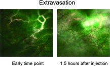
| The behavior of targeted quantum dots (Qdots) across three different tumor models using intravital microscopy with submicrometer resolution is described. As in the figure, the differences in extravasation between tumor types are shown and the kinetics are quatnified. Further, by demonstrating similarity in Qdot binding to tumor blood vessels across different tumor types, this work suggests several advantages implicit in vascular targeting compared with tumor cell targeting. |
Abstract:
Though targeted nanoparticle-based imaging agents and therapeutics for diagnosing and treating cancer are making their way to and through the clinical trials process, researchers still do not have a good understanding of how nanoparticles reach tumors and how they then bind to and enter the targeted tumor. To overcome that knowledge deficit, two teams of investigators, both part of the Alliance for Nanotechnology in Cancer have undertaken studies aiming to track nanoparticles as they move through living animals.
Tracking Tumor-Targeting Nanoparticles in the Body
Bethesda, MD | Posted on October 27th, 2010In one study, a team of investigators at Stanford University used quantum dots to study how nanoparticles travel through tumor blood vessels in living test subjects, bind to molecular targets on the surface of those blood vessels, and then travel out of the blood stream and into the tumor itself. Sanjiv Sam Gambhir, co-director of one of nine National Cancer Institute (NCI) Centers of Cancer Nanotechnology Excellence, led this study. He and his colleagues published their findings in the journal Small. In a second study, published in the journal ACS Nano, Alliance investigators Dong Shin, Mostafa El-Sayed, and Shuming Nie of Emory University and the Georgia Institute of Technology used targeted gold nanocrystals to study both active and passive targeting of tumors.
In the Stanford study, Dr. Gambhir and his collaborators exploited the capabilities of intravital microscopy, a technique that enables researchers to see brightly fluorescent markers through a living animal's skin in real time. In this series of experiments, the Stanford team examined nanoparticle trafficking in mice in which a variety of different types of tumors were allowed to grow in the animals' ears. For the fluorescent marker, the investigators used a near-infrared emitting quantum dot linked to RGD, a molecule known to bind tightly to a protein found on the surface of blood vessels surrounding tumors.
To their surprise, the researchers found that regardless of the type of tumor studied, nanoparticle binding only occurred when aggregates of particles - not single particles - were able to tether themselves to multiple, discreet sites within a tumor. The researchers were not able to detect any significant binding when they repeated these experiments using quantum dots lacking the RGD targeting molecule. The investigators also found that binding rates and binding patterns were consistent across all tumor types, a reassuring finding given the natural heterogeneity that characterizes human cancers.
While binding ability appears to be independent of tumor type, the same cannot be said for extravasation, i.e., the transit of a nanoparticle out of the blood stream and into a tumor. The researchers noted in their paper that it is likely that nanoparticle shape and size will play a critical role in determining how a given nanoparticle will extravasate into each particular type of tumor.
Meanwhile, the Emory-Georgia Tech team used rod-shaped gold nanocrystals linked to tumor-targeting peptides to explore the delivery mechanisms that enable nanoparticles to accumulate in tumors. The investigators used gold nanoparticles so that they could quantify the number of nanoparticles reaching tumors and other tissues. Gold does not occur naturally in mammals, so any gold detected in a given tumor or tissue using the highly sensitive and accurate technique known as elemental mass spectrometry would had to have come from gold nanoparticles.
To conduct their experiments, the investigators created three formulations by attaching one of three tumor-targeting molecules to the surface of the gold nanorods. They then injected the nanoparticles into animals bearing implanted human tumors, allowed the nanoparticles to circulate through the body, and measured the amount of gold that accumulated in the implanted tumors and other tissues. The researchers also repeated this experiment using untargeted gold nanoparticles. The results were surprising in that the targeting molecules only marginally increased the amount of gold that accumulated in tumors.
The investigators concluded that gold nanoparticles designed to be used in photothermal anticancer therapy should be injected directly into tumors rather than via intravenous administration in order to achieve the greatest concentration of gold in tumors. They also noted in their paper that these experiments suggest that target binding is not the rate limiting step for nanoparticle delivery, but rather that transport out of the blood stream and into tumors is the major barrier to nanoparticle accumulation in tumors.
The work using intravital microscopy, which is detailed in a paper titled, "Dynamic Visualization of RGD-Quantum Dot Binding to Tumor Neovasculature and Extravasation in Multiple Living Mouse Models Using Intravital Microscopy," was supported in part by the NCI Alliance for Nanotechnology in Cancer, a comprehensive initiative designed to accelerate the application of nanotechnology to the prevention, diagnosis, and treatment of cancer. An abstract of this paper is available at the journal's website.
View Abstract at dx.doi.org/doi:10.1002/smll.201001022
The work using gold nanocrystals, which is detailed in a paper titled, "A Reexamination of Active and Passive Tumor Targeting by Using Rod-Shaped Gold Nanocrystals and Covalently Targeted Peptide Ligands," was also supported in part by the NCI Alliance for Nanotechnology in Cancer. An abstract of this paper is available at the journal's website.
View abstract at pubs.acs.org/doi/abs/10.1021/nn102055s
####
About NCI Alliance for Nanotechnology in Cancer
To help meet the goal of reducing the burden of cancer, the National Cancer Institute (NCI), part of the National Institutes of Health, is engaged in efforts to harness the power of nanotechnology to radically change the way we diagnose, treat and prevent cancer.
The NCI Alliance for Nanotechnology in Cancer is a comprehensive, systematized initiative encompassing the public and private sectors, designed to accelerate the application of the best capabilities of nanotechnology to cancer.
Currently, scientists are limited in their ability to turn promising molecular discoveries into benefits for cancer patients. Nanotechnology can provide the technical power and tools that will enable those developing new diagnostics, therapeutics, and preventives to keep pace with today’s explosion in knowledge.
For more information, please click here
Copyright © NCI Alliance for Nanotechnology in Cancer
If you have a comment, please Contact us.Issuers of news releases, not 7th Wave, Inc. or Nanotechnology Now, are solely responsible for the accuracy of the content.
| Related News Press |
News and information
![]() Decoding hydrogen‑bond network of electrolyte for cryogenic durable aqueous zinc‑ion batteries January 30th, 2026
Decoding hydrogen‑bond network of electrolyte for cryogenic durable aqueous zinc‑ion batteries January 30th, 2026
![]() COF scaffold membrane with gate‑lane nanostructure for efficient Li+/Mg2+ separation January 30th, 2026
COF scaffold membrane with gate‑lane nanostructure for efficient Li+/Mg2+ separation January 30th, 2026
Govt.-Legislation/Regulation/Funding/Policy
![]() Metasurfaces smooth light to boost magnetic sensing precision January 30th, 2026
Metasurfaces smooth light to boost magnetic sensing precision January 30th, 2026
![]() New imaging approach transforms study of bacterial biofilms August 8th, 2025
New imaging approach transforms study of bacterial biofilms August 8th, 2025
![]() Electrifying results shed light on graphene foam as a potential material for lab grown cartilage June 6th, 2025
Electrifying results shed light on graphene foam as a potential material for lab grown cartilage June 6th, 2025
Academic/Education
![]() Rice University launches Rice Synthetic Biology Institute to improve lives January 12th, 2024
Rice University launches Rice Synthetic Biology Institute to improve lives January 12th, 2024
![]() Multi-institution, $4.6 million NSF grant to fund nanotechnology training September 9th, 2022
Multi-institution, $4.6 million NSF grant to fund nanotechnology training September 9th, 2022
Nanomedicine
![]() New molecular technology targets tumors and simultaneously silences two ‘undruggable’ cancer genes August 8th, 2025
New molecular technology targets tumors and simultaneously silences two ‘undruggable’ cancer genes August 8th, 2025
![]() New imaging approach transforms study of bacterial biofilms August 8th, 2025
New imaging approach transforms study of bacterial biofilms August 8th, 2025
![]() Cambridge chemists discover simple way to build bigger molecules – one carbon at a time June 6th, 2025
Cambridge chemists discover simple way to build bigger molecules – one carbon at a time June 6th, 2025
![]() Electrifying results shed light on graphene foam as a potential material for lab grown cartilage June 6th, 2025
Electrifying results shed light on graphene foam as a potential material for lab grown cartilage June 6th, 2025
Announcements
![]() Decoding hydrogen‑bond network of electrolyte for cryogenic durable aqueous zinc‑ion batteries January 30th, 2026
Decoding hydrogen‑bond network of electrolyte for cryogenic durable aqueous zinc‑ion batteries January 30th, 2026
![]() COF scaffold membrane with gate‑lane nanostructure for efficient Li+/Mg2+ separation January 30th, 2026
COF scaffold membrane with gate‑lane nanostructure for efficient Li+/Mg2+ separation January 30th, 2026
Quantum Dots/Rods
![]() A new kind of magnetism November 17th, 2023
A new kind of magnetism November 17th, 2023
![]() IOP Publishing celebrates World Quantum Day with the announcement of a special quantum collection and the winners of two prestigious quantum awards April 14th, 2023
IOP Publishing celebrates World Quantum Day with the announcement of a special quantum collection and the winners of two prestigious quantum awards April 14th, 2023
![]() Qubits on strong stimulants: Researchers find ways to improve the storage time of quantum information in a spin rich material January 27th, 2023
Qubits on strong stimulants: Researchers find ways to improve the storage time of quantum information in a spin rich material January 27th, 2023
![]() NIST’s grid of quantum islands could reveal secrets for powerful technologies November 18th, 2022
NIST’s grid of quantum islands could reveal secrets for powerful technologies November 18th, 2022
Nanobiotechnology
![]() New molecular technology targets tumors and simultaneously silences two ‘undruggable’ cancer genes August 8th, 2025
New molecular technology targets tumors and simultaneously silences two ‘undruggable’ cancer genes August 8th, 2025
![]() New imaging approach transforms study of bacterial biofilms August 8th, 2025
New imaging approach transforms study of bacterial biofilms August 8th, 2025
![]() Ben-Gurion University of the Negev researchers several steps closer to harnessing patient's own T-cells to fight off cancer June 6th, 2025
Ben-Gurion University of the Negev researchers several steps closer to harnessing patient's own T-cells to fight off cancer June 6th, 2025
![]() Electrifying results shed light on graphene foam as a potential material for lab grown cartilage June 6th, 2025
Electrifying results shed light on graphene foam as a potential material for lab grown cartilage June 6th, 2025
Research partnerships
![]() Lab to industry: InSe wafer-scale breakthrough for future electronics August 8th, 2025
Lab to industry: InSe wafer-scale breakthrough for future electronics August 8th, 2025
![]() HKU physicists uncover hidden order in the quantum world through deconfined quantum critical points April 25th, 2025
HKU physicists uncover hidden order in the quantum world through deconfined quantum critical points April 25th, 2025
|
|
||
|
|
||
| The latest news from around the world, FREE | ||
|
|
||
|
|
||
| Premium Products | ||
|
|
||
|
Only the news you want to read!
Learn More |
||
|
|
||
|
Full-service, expert consulting
Learn More |
||
|
|
||


