Home > Press > DNA detection: Color by nanoparticles
 |
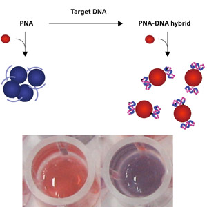
| Fig. 1: Schematic diagram showing the conversion of PNA to a PNA–DNA complex (top) and photographs (bottom) of gold nanoparticle solutions with the addition of a PNA–DNA complex (left) and PNA (right). © 2009 A*STAR |
Abstract:
Using a precise and fast nanoparticle-based technique, DNA detection by the naked eye is now possible
DNA detection: Color by nanoparticles
Singapore | Posted on December 23rd, 2009A sensitive yet uncomplicated method to detect differences in DNA strands using metal nanoparticle solutions has been developed by Roejarek Kanjanawarut and Xiaodi Su at the Institute of Materials Research and Engineering at A*STAR, Singapore (1). The method requires no modification of the surfaces of the nanoparticles, making it particularly fast and versatile to implement.
The researchers worked on the principle that aggregated and dispersed nanoparticles have different optical properties that make the solutions take on different colors. This means that the results of their tests can be displayed in minutes, and recorded qualitatively by the naked eye and quantitatively by a standard spectrometer.
The tendency of nanoparticles to aggregate in solution has been considered a drawback, and in previous approaches to use them for sensing, DNA strands were directly attached to the particles' surfaces to prevent them clumping together. Kanjanawarut and Su turned the tables to take advantage of the natural propensity to aggregate as inspiration for their assays. Compared with the earlier nanoparticle- and chip-based DNA assays, the new method is cost-effective as it involves no time-consuming surface modifications, particle bioconjugation, biohazardous labeling or tedious assay procedures, explains Su.
The researchers used gold and silver nanoparticles that have a coating of negatively charged molecules. These charges repel one another, preventing the nanoparticles from aggregating in solution. They then added peptide nucleic acid (PNA) probes to the solution, which bound to the negative surfaces, shielding the charge and allowing the nanoparticles to agglomerate. The solution with gold nanoparticles changed from bright red to dark purple.
The PNA can be tailored to bind selectively to whichever DNA strand is the target for detection. After adding the target DNA strand to the solution, it bound to the PNA, making a PNA-DNA complex. The solution remained bright red, clearly different from the aggregated dark purple solution (Fig. 1). Even one mismatched DNA base compared to the target strand could be detected.
The color difference results from the formation of the PNA-DNA complex on the nanoparticle surface, which is negatively charged due to the negative charge born by the DNA chains; this makes the nanoparticles repel one another and thus take longer to aggregate.
Su hopes that their approach can be used to measure specific DNA sequences for diagnosis or fundamental research. "The sequence involved in this study, for example, is from a human gene. Single-base-mismatch detection in this gene is associated with, and thus markers for, autoimmune diseases such as psoriasis," she says.
The A*STAR affiliated authors on this highlight are from the Institute of Materials Research and Engineering (IMRE) (www.imre.a-star.edu.sg/)
Reference
1. Kanjanawarut, R. & Su, X. Colorimetric detection of DNA using unmodified metallic nanoparticles and peptide nucleic acid probes. Analytical Chemistry 81, 6122-6129 (2009).
####
About A*STAR Research
A*STAR Research is an online and print publication highlighting some of the best research and technological developments at the research institutes of Singapore’s Agency for Science, Technology and Research (A*STAR). Established in 2002, A*STAR has thrived as a global research organization with a principal mission of fostering world-class scientific research and talent for a vibrant knowledge-based Singapore. A*STAR currently oversees 14 research institutes as well as 7 consortia and centers located in the Biopolis and Fusionopolis complexes and the vicinity, and supports extramural research in collaboration with universities, hospital research centers and other local and international partners. The various A*STAR institutes are involved in research in a wide range of scientific fields, coordinated and funded by Singapore’s Biomedical Research Council (BMRC) and Science and Engineering Research Council (SERC).
For more information, please click here
Copyright © A*STAR Research
If you have a comment, please Contact us.Issuers of news releases, not 7th Wave, Inc. or Nanotechnology Now, are solely responsible for the accuracy of the content.
| Related News Press |
News and information
![]() Researchers develop molecular qubits that communicate at telecom frequencies October 3rd, 2025
Researchers develop molecular qubits that communicate at telecom frequencies October 3rd, 2025
![]() Next-generation quantum communication October 3rd, 2025
Next-generation quantum communication October 3rd, 2025
![]() "Nanoreactor" cage uses visible light for catalytic and ultra-selective cross-cycloadditions October 3rd, 2025
"Nanoreactor" cage uses visible light for catalytic and ultra-selective cross-cycloadditions October 3rd, 2025
Nanomedicine
![]() New molecular technology targets tumors and simultaneously silences two ‘undruggable’ cancer genes August 8th, 2025
New molecular technology targets tumors and simultaneously silences two ‘undruggable’ cancer genes August 8th, 2025
![]() New imaging approach transforms study of bacterial biofilms August 8th, 2025
New imaging approach transforms study of bacterial biofilms August 8th, 2025
![]() Cambridge chemists discover simple way to build bigger molecules – one carbon at a time June 6th, 2025
Cambridge chemists discover simple way to build bigger molecules – one carbon at a time June 6th, 2025
![]() Electrifying results shed light on graphene foam as a potential material for lab grown cartilage June 6th, 2025
Electrifying results shed light on graphene foam as a potential material for lab grown cartilage June 6th, 2025
Announcements
![]() Rice membrane extracts lithium from brines with greater speed, less waste October 3rd, 2025
Rice membrane extracts lithium from brines with greater speed, less waste October 3rd, 2025
![]() Researchers develop molecular qubits that communicate at telecom frequencies October 3rd, 2025
Researchers develop molecular qubits that communicate at telecom frequencies October 3rd, 2025
![]() Next-generation quantum communication October 3rd, 2025
Next-generation quantum communication October 3rd, 2025
![]() "Nanoreactor" cage uses visible light for catalytic and ultra-selective cross-cycloadditions October 3rd, 2025
"Nanoreactor" cage uses visible light for catalytic and ultra-selective cross-cycloadditions October 3rd, 2025
Nanobiotechnology
![]() New molecular technology targets tumors and simultaneously silences two ‘undruggable’ cancer genes August 8th, 2025
New molecular technology targets tumors and simultaneously silences two ‘undruggable’ cancer genes August 8th, 2025
![]() New imaging approach transforms study of bacterial biofilms August 8th, 2025
New imaging approach transforms study of bacterial biofilms August 8th, 2025
![]() Ben-Gurion University of the Negev researchers several steps closer to harnessing patient's own T-cells to fight off cancer June 6th, 2025
Ben-Gurion University of the Negev researchers several steps closer to harnessing patient's own T-cells to fight off cancer June 6th, 2025
![]() Electrifying results shed light on graphene foam as a potential material for lab grown cartilage June 6th, 2025
Electrifying results shed light on graphene foam as a potential material for lab grown cartilage June 6th, 2025
|
|
||
|
|
||
| The latest news from around the world, FREE | ||
|
|
||
|
|
||
| Premium Products | ||
|
|
||
|
Only the news you want to read!
Learn More |
||
|
|
||
|
Full-service, expert consulting
Learn More |
||
|
|
||


