Home > Press > 'Label-free' imaging tool tracks nanotubes in cells, blood for biomedical research
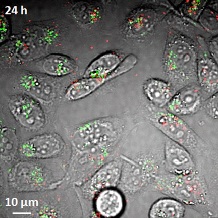 |
| Researchers have demonstrated a new imaging tool for tracking structures called single-wall carbon nanotubes in living cells and the bloodstream, work that could aid efforts to perfect their use in laboratory or medical applications. Here, the imaging system detects both metallic and semiconducting nanotubes, false-colored in red and green, in live hamster cells. (Weldon School of Biomedical Engineering, Purdue University) |
Abstract:
Label-Free Imaging of Semiconducting and Metallic Carbon Nanotubes in Cells and Mice Using Transient Absorption Microscopy
Ling Tong1, Yuxiang Liu2, Bridget D. Dolash3, Yookyung Jung4, Mikhail N. Slipchenko2, Donald E. Bergstrom3,5 and Ji-Xin Cheng1,2,5*
1Department of Chemistry; 2Weldon School of Biomedical Engineering, 3Department of Medical Chemistry and Molecular Pharmacology; 4Department of Physics,5 Birck Nanotechnology Center, Purdue University
As interest in the potential biomedical applications of carbon nanotubes increases, there is a need for methods that can image nanotubes in live cells, tissues and animals. Although techniques such as Raman, photoacoustic and near-infrared photoluminescence imaging have been used to visualize nanotubes in biological environments, these techniques are limited because nanotubes provide only weak photoluminescence and low Raman scattering and it remains difficult to image both semiconducting and metallic nanotubes at the same time. Here, we show that transient absorption microscopy offers a label-free method to image both semiconducting and metallic single-walled carbon nanotubes in vitro and in vivo, in real time, with submicrometre resolution. By using appropriate near-infrared excitation wavelengths, we detect strong transient absorption signals with opposite phases from semiconducting and metallic nanotubes. Our method separates background signals generated by red blood cells and this allows us to follow the movement of both types of nanotubes inside cells and in the blood circulation and organs of mice without any significant damaging effects.
'Label-free' imaging tool tracks nanotubes in cells, blood for biomedical research
West Lafayette, IN | Posted on December 5th, 2011Researchers have demonstrated a new imaging tool for tracking structures called carbon nanotubes in living cells and the bloodstream, which could aid efforts to perfect their use in biomedical research and clinical medicine.
The structures have potential applications in drug delivery to treat diseases and imaging for cancer research. Two types of nanotubes are created in the manufacturing process, metallic and semiconducting. Until now, however, there has been no technique to see both types in living cells and the bloodstream, said Ji-Xin Cheng, an associate professor of biomedical engineering and chemistry at Purdue University.
The imaging technique, called transient absorption, uses a pulsing near-infrared laser to deposit energy into the nanotubes, which then are probed by a second near-infrared laser.
The researchers have overcome key obstacles in using the imaging technology, detecting and monitoring the nanotubes in live cells and laboratory mice, Cheng said.
"Because we can do this at high speed, we can see what's happening in real time as the nanotubes are circulating in the bloodstream," he said.
Findings are detailed in a research paper posted online Sunday (Dec. 4) in the journal Nature Nanotechnology.
The imaging technique is "label free," meaning it does not require that the nanotubes be marked with dyes, making it potentially practical for research and medicine, Cheng said.
"It's a fundamental tool for research that will provide information for the scientific community to learn how to perfect the use of nanotubes for biomedical and clinical applications," he said.
The conventional imaging method uses luminescence, which is limited because it detects the semiconducting nanotubes but not the metallic ones.
The nanotubes have a diameter of about 1 nanometer, or roughly the length of 10 hydrogen atoms strung together, making them far too small to be seen with a conventional light microscope. One challenge in using the transient absorption imaging system for living cells was to eliminate the interference caused by the background glow of red blood cells, which is brighter than the nanotubes.
The researchers solved this problem by separating the signals from red blood cells and nanotubes in two separate "channels." Light from the red blood cells is slightly delayed compared to light emitted by the nanotubes. The two types of signals are "phase separated" by restricting them to different channels based on this delay.
Researchers used the technique to see nanotubes circulating in the blood vessels of mice earlobes.
"This is important for drug delivery because you want to know how long nanotubes remain in blood vessels after they are injected," Cheng said. "So you need to visualize them in real time circulating in the bloodstream."
The structures, called single-wall carbon nanotubes, are formed by rolling up a one-atom-thick layer of graphite called graphene. The nanotubes are inherently hydrophobic, so some of the nanotubes used in the study were coated with DNA to make them water-soluble, which is required for them to be transported in the bloodstream and into cells.
The researchers also have taken images of nanotubes in the liver and other organs to study their distribution in mice, and they are using the imaging technique to study other nanomaterials such as graphene.
The paper was written by doctoral student Ling Tong; postdoctoral research associate Yuxiang Liu; doctoral students Bridget D. Dolash and Yookyung Jung; biomedical engineering research scientist Mikhail N. Slipchenko; Donald E. Bergstrom, the Walther Professor of Medicinal Chemistry; and Cheng.
The research is funded by the National Science Foundation.
####
For more information, please click here
Contacts:
Writer:
Emil Venere
765-494-4709
Source:
Ji-Xin Cheng
765-494-4335
Copyright © Purdue University
If you have a comment, please Contact us.Issuers of news releases, not 7th Wave, Inc. or Nanotechnology Now, are solely responsible for the accuracy of the content.
Bookmark:


| Related Links |
![]() Purdue Weldon School of Biomedical Engineering
Purdue Weldon School of Biomedical Engineering
![]() Purdue Department of Chemistry
Purdue Department of Chemistry
| Related News Press |
News and information
![]() Quantum computer improves AI predictions April 17th, 2026
Quantum computer improves AI predictions April 17th, 2026
![]() Flexible sensor gains sensitivity under pressure April 17th, 2026
Flexible sensor gains sensitivity under pressure April 17th, 2026
![]() A reusable chip for particulate matter sensing April 17th, 2026
A reusable chip for particulate matter sensing April 17th, 2026
![]() Detecting vibrational quantum beating in the predissociation dynamics of SF6 using time-resolved photoelectron spectroscopy April 17th, 2026
Detecting vibrational quantum beating in the predissociation dynamics of SF6 using time-resolved photoelectron spectroscopy April 17th, 2026
Imaging
![]() Simple algorithm paired with standard imaging tool could predict failure in lithium metal batteries August 8th, 2025
Simple algorithm paired with standard imaging tool could predict failure in lithium metal batteries August 8th, 2025
Graphene/ Graphite
![]() Electrifying results shed light on graphene foam as a potential material for lab grown cartilage June 6th, 2025
Electrifying results shed light on graphene foam as a potential material for lab grown cartilage June 6th, 2025
![]() Breakthrough in proton barrier films using pore-free graphene oxide: Kumamoto University researchers achieve new milestone in advanced coating technologies September 13th, 2024
Breakthrough in proton barrier films using pore-free graphene oxide: Kumamoto University researchers achieve new milestone in advanced coating technologies September 13th, 2024
Govt.-Legislation/Regulation/Funding/Policy
![]() Quantum computer improves AI predictions April 17th, 2026
Quantum computer improves AI predictions April 17th, 2026
![]() Metasurfaces smooth light to boost magnetic sensing precision January 30th, 2026
Metasurfaces smooth light to boost magnetic sensing precision January 30th, 2026
![]() New imaging approach transforms study of bacterial biofilms August 8th, 2025
New imaging approach transforms study of bacterial biofilms August 8th, 2025
Chip Technology
![]() A reusable chip for particulate matter sensing April 17th, 2026
A reusable chip for particulate matter sensing April 17th, 2026
![]() Metasurfaces smooth light to boost magnetic sensing precision January 30th, 2026
Metasurfaces smooth light to boost magnetic sensing precision January 30th, 2026
Nanotubes/Buckyballs/Fullerenes/Nanorods/Nanostrings/Nanosheets
![]() Tiny nanosheets, big leap: A new sensor detects ethanol at ultra-low levels January 30th, 2026
Tiny nanosheets, big leap: A new sensor detects ethanol at ultra-low levels January 30th, 2026
![]() Enhancing power factor of p- and n-type single-walled carbon nanotubes April 25th, 2025
Enhancing power factor of p- and n-type single-walled carbon nanotubes April 25th, 2025
![]() Chainmail-like material could be the future of armor: First 2D mechanically interlocked polymer exhibits exceptional flexibility and strength January 17th, 2025
Chainmail-like material could be the future of armor: First 2D mechanically interlocked polymer exhibits exceptional flexibility and strength January 17th, 2025
![]() Innovative biomimetic superhydrophobic coating combines repair and buffering properties for superior anti-erosion December 13th, 2024
Innovative biomimetic superhydrophobic coating combines repair and buffering properties for superior anti-erosion December 13th, 2024
Nanomedicine
![]() A fundamentally new therapeutic approach to cystic fibrosis: Nanobody repairs cellular defect April 17th, 2026
A fundamentally new therapeutic approach to cystic fibrosis: Nanobody repairs cellular defect April 17th, 2026
![]() New molecular technology targets tumors and simultaneously silences two �undruggable� cancer genes August 8th, 2025
New molecular technology targets tumors and simultaneously silences two �undruggable� cancer genes August 8th, 2025
![]() New imaging approach transforms study of bacterial biofilms August 8th, 2025
New imaging approach transforms study of bacterial biofilms August 8th, 2025
![]() Electrifying results shed light on graphene foam as a potential material for lab grown cartilage June 6th, 2025
Electrifying results shed light on graphene foam as a potential material for lab grown cartilage June 6th, 2025
Discoveries
![]() Quantum computer improves AI predictions April 17th, 2026
Quantum computer improves AI predictions April 17th, 2026
![]() Flexible sensor gains sensitivity under pressure April 17th, 2026
Flexible sensor gains sensitivity under pressure April 17th, 2026
![]() A reusable chip for particulate matter sensing April 17th, 2026
A reusable chip for particulate matter sensing April 17th, 2026
![]() Detecting vibrational quantum beating in the predissociation dynamics of SF6 using time-resolved photoelectron spectroscopy April 17th, 2026
Detecting vibrational quantum beating in the predissociation dynamics of SF6 using time-resolved photoelectron spectroscopy April 17th, 2026
Announcements
![]() A fundamentally new therapeutic approach to cystic fibrosis: Nanobody repairs cellular defect April 17th, 2026
A fundamentally new therapeutic approach to cystic fibrosis: Nanobody repairs cellular defect April 17th, 2026
![]() UC Irvine physicists discover method to reverse �quantum scrambling� : The work addresses the problem of information loss in quantum computing system April 17th, 2026
UC Irvine physicists discover method to reverse �quantum scrambling� : The work addresses the problem of information loss in quantum computing system April 17th, 2026
Tools
![]() Metasurfaces smooth light to boost magnetic sensing precision January 30th, 2026
Metasurfaces smooth light to boost magnetic sensing precision January 30th, 2026
![]() From sensors to smart systems: the rise of AI-driven photonic noses January 30th, 2026
From sensors to smart systems: the rise of AI-driven photonic noses January 30th, 2026
![]() Japan launches fully domestically produced quantum computer: Expo visitors to experience quantum computing firsthand August 8th, 2025
Japan launches fully domestically produced quantum computer: Expo visitors to experience quantum computing firsthand August 8th, 2025
Safety-Nanoparticles/Risk management
![]() Tiny nanosheets, big leap: A new sensor detects ethanol at ultra-low levels January 30th, 2026
Tiny nanosheets, big leap: A new sensor detects ethanol at ultra-low levels January 30th, 2026
![]() Onion-like nanoparticles found in aircraft exhaust May 14th, 2025
Onion-like nanoparticles found in aircraft exhaust May 14th, 2025
![]() Closing the gaps � MXene-coating filters can enhance performance and reusability February 28th, 2025
Closing the gaps � MXene-coating filters can enhance performance and reusability February 28th, 2025
Photonics/Optics/Lasers
![]() Metasurfaces smooth light to boost magnetic sensing precision January 30th, 2026
Metasurfaces smooth light to boost magnetic sensing precision January 30th, 2026
![]() From sensors to smart systems: the rise of AI-driven photonic noses January 30th, 2026
From sensors to smart systems: the rise of AI-driven photonic noses January 30th, 2026
|
|
||
|
|
||
| The latest news from around the world, FREE | ||
|
|
||
|
|
||
| Premium Products | ||
|
|
||
|
Only the news you want to read!
Learn More |
||
|
|
||
|
Full-service, expert consulting
Learn More |
||
|
|
||