Home > Press > Nano-probes Allow an Inside Look at Cell Nuclei
Abstract:
Biologists may soon use it to watch the inner workings of a living cell like never before
Nano-probes Allow an Inside Look at Cell Nuclei
Berkeley, CA | March 18, 2005
Nanotechnology may be in its infancy, but biologists may soon use it to watch the inner workings of a living cell like never before. Scientists at the U.S. Department of Energy’s Lawrence Berkeley National Laboratory (Berkeley Lab) have developed a way to sneak nano-sized probes inside cell nuclei where they can track life’s fundamental processes, such as DNA repair, for hours on end.
“Our work represents the first time a biologist can image long-term phenomena within the nuclei of living cells,” says Fanqing Chen of Berkeley Lab’s Life Sciences Division, who developed the technique with Daniele Gerion of Lawrence Livermore National Laboratory.

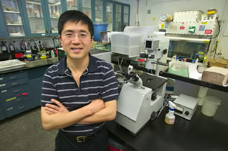
Fanqing Chen (pictured) and Daniele Gerion have harnessed the powers of nanotechnology to image the interior of cell nuclei.
|
Their success lies in specially prepared crystalline semiconductors composed of a few hundred or thousand atoms that emit different colors of light when illuminated by a laser. Because these fluorescent probes are stable and nontoxic, they have the ability to remain in a cell’s nucleus — without harming the cell or fading out — much longer than conventional fluorescent labels. This could give biologists a ringside seat to nuclear processes that span several hours or days, such as DNA replication, genomic alterations, and cell cycle control. The long-lived probes may also allow researchers to track the effectiveness of disease-fighting drugs that target these processes.
“We could determine whether a drug has arrived where it is supposed to, and if it is having the desired impact,” says Chen.
The first enduring look into the secret lives of cell nuclei comes by way of a strong collaboration between biologists and chemists. For the past four years, Chen and Gerion have worked closely with members of the lab of Paul Alivisatos, a Berkeley Lab chemist in the Materials Sciences Division and Associate Laboratory Director who helped pioneer the development of nano-sized crystals of semiconductor materials. Called quantum dots, these microscopic crystals have shown promise in such wide-ranging applications as solar cells, computer design, and biology. In 1998, for example, Alivisatos developed a way to fashion inorganic nanocrystals composed of cadmium selenide and cadmium sulfide into fluorescent probes suitable for the study of living cells. This technology has been licensed to the Hayward, California-based Quantum Dot Corporation for use in biological assays.
More recently, Chen and Gerion wondered if they could get even closer to the genetic action by transporting quantum dots inside cell nuclei.
“We took the tool Paul developed and applied it to a problem faced by biologists every day — getting inside the nucleus, a desirable target because the cell’s genetic information resides there,” says Chen.

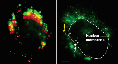
These two images portray the movement of the nano-sized probes. On the left, a false-color overlay of fluorescence from a cell taken at four minute intervals reveals the dots moving from the green to the red positions. On the right, a large aggregate of immobile dots is indicated with the red arrow, while the circled stars and arrows indicate dots that move.
|
First, they had to breach the nuclear membrane, which has pores that are only about 20 nanometers wide. To fit through these tiny slits, Chen and Gerion used an especially compact cadmium selenide/zinc sulfide quantum dot coated with silica. Next, they stole a trick from a virus’s playbook to smuggle this nanocrystal past the highly selective membrane that guards the entrance into the nucleus. In nature, a virus called SV40 is coated with a protein that binds to a cell’s nuclear trafficking mechanism, a ploy that gives the virus an unhindered ride inside the nucleus. Chen and Gerion obtained a portion of this protein and attached it to the quantum dot. The result is a hybrid quantum dot, part biological molecule and part nano-sized semiconductor, that is small enough to slide through the nuclear membrane’s pores and believable enough to slip past the membrane’s barriers.
“We knew we could get quantum dots inside a cell, but getting them through the nuclear membrane is very difficult,” says Chen. “So we learned from the virus.”
So far, Chen and Gerion have been able to introduce and retain quantum dots in the nuclei of living cells for up to a week without harming the cell. In addition, quantum dots fluoresce for days at a resolution high enough to detect biological events carried out by single molecules. In contrast, conventional labels such as organic fluorescent dyes and green fluorescent proteins only fluoresce for a few minutes at a high resolution. These labels are also either toxic to cells or difficult to construct and manipulate.
In the future, they hope to tailor quantum dots to track specific chemical reactions inside nuclei, such as how proteins help repair DNA after irradiation. They have already visualized the dots’ journey from the area surrounding the nucleus to inside the nucleus, a feat that opens the door for real-time observations of nuclear trafficking mechanisms. They also hope to target other cellular organelles besides the nucleus, such as mitochondria and Golgi bodies. And because quantum dots emit different colors of light based on their size, they can be used to observe the transfer of material between cells.
“We can have two different quantum dots in two different cells, and watch as the cells exchange their mitochondria,” says Chen, adding that their technique paves the way for imaging a host of other long-term biological events. “The toughest part is getting inside the nucleus, and we have already cleared that hurdle.”
Chen and Gerion’s research was published in the 2004, Vol. 2, No. 10 issue of Nano Letters.
About Berkeley Lab
Berkeley Lab is a U.S. Department of Energy national laboratory located in Berkeley, California. It conducts unclassified scientific research and is managed by the University of California. Visit our Website at www.lbl.gov.
Contact:
Dan Krotz
(510) 486-4019
dakrotz@lbl.gov
Copyright © Berkeley Lab
If you have a comment, please Contact us.
Issuers of news releases, not 7th Wave, Inc. or Nanotechnology Now, are solely responsible for the accuracy of the content.
| Related News Press |
Possible Futures
![]() Spinel-type sulfide semiconductors to operate the next-generation LEDs and solar cells For solar-cell absorbers and green-LED source October 3rd, 2025
Spinel-type sulfide semiconductors to operate the next-generation LEDs and solar cells For solar-cell absorbers and green-LED source October 3rd, 2025
Nanomedicine
![]() New molecular technology targets tumors and simultaneously silences two ‘undruggable’ cancer genes August 8th, 2025
New molecular technology targets tumors and simultaneously silences two ‘undruggable’ cancer genes August 8th, 2025
![]() New imaging approach transforms study of bacterial biofilms August 8th, 2025
New imaging approach transforms study of bacterial biofilms August 8th, 2025
![]() Cambridge chemists discover simple way to build bigger molecules – one carbon at a time June 6th, 2025
Cambridge chemists discover simple way to build bigger molecules – one carbon at a time June 6th, 2025
![]() Electrifying results shed light on graphene foam as a potential material for lab grown cartilage June 6th, 2025
Electrifying results shed light on graphene foam as a potential material for lab grown cartilage June 6th, 2025
Announcements
![]() Rice membrane extracts lithium from brines with greater speed, less waste October 3rd, 2025
Rice membrane extracts lithium from brines with greater speed, less waste October 3rd, 2025
![]() Researchers develop molecular qubits that communicate at telecom frequencies October 3rd, 2025
Researchers develop molecular qubits that communicate at telecom frequencies October 3rd, 2025
![]() Next-generation quantum communication October 3rd, 2025
Next-generation quantum communication October 3rd, 2025
![]() "Nanoreactor" cage uses visible light for catalytic and ultra-selective cross-cycloadditions October 3rd, 2025
"Nanoreactor" cage uses visible light for catalytic and ultra-selective cross-cycloadditions October 3rd, 2025
Tools
![]() Japan launches fully domestically produced quantum computer: Expo visitors to experience quantum computing firsthand August 8th, 2025
Japan launches fully domestically produced quantum computer: Expo visitors to experience quantum computing firsthand August 8th, 2025
![]() Rice researchers harness gravity to create low-cost device for rapid cell analysis February 28th, 2025
Rice researchers harness gravity to create low-cost device for rapid cell analysis February 28th, 2025
|
|
||
|
|
||
| The latest news from around the world, FREE | ||
|
|
||
|
|
||
| Premium Products | ||
|
|
||
|
Only the news you want to read!
Learn More |
||
|
|
||
|
Full-service, expert consulting
Learn More |
||
|
|
||


