Home > Press > Electron tomography with 3,487 images in 3.5 seconds: High-speed electron tomography sets new standards for 3-D images of the nanoworld
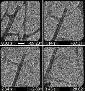 |
| This image series serves as a data basis for the reconstruction of the 3-D electron tomogram.
Copyright: Migunov, V. et al. Sci. Rep. 5, 14516, 2015 (CC BY 4.0) |
Abstract:
Scientists from the Ernst Ruska-Centre in Forschungszentrum Jülich used a transmission electron microscope to record almost 3500 images in 3.5 seconds for the reconstruction of a 3D electron tomogram. Previously, 10 to 60 minutes and a ten-fold greater electron dose were required to record such image sequences. The new capability is particularly suitable for examining biological cells, bacteria and viruses, whose structure can be damaged by the electron beam. In addition, it enables dynamic processes, such as chemical reactions and electronic switching phenomena, to be visualized in real time in three dimensions with sub-nanometre precision. The findings have been published in the journal Scientific Reports.
Electron tomography with 3,487 images in 3.5 seconds: High-speed electron tomography sets new standards for 3-D images of the nanoworld
Jülich, Germany | Posted on October 6th, 2015Electron tomography is related to computed tomography, which has become indispensable in research and clinical studies. Electron tomograms can be obtained from much smaller volumes than with X-ray-based techniques. The three dimensional spatial resolution of electron tomography is the highest achievable with today's technology. The method is uniquely suited for studying viruses and bacteria to facilitate development of medications, or for imaging the structures of novel nanomaterials for applications that range from nanoelectronics to energy technology.
"The ability to accelerate image acquisition and reduce radiation dose opens up new horizons, particularly in life sciences and soft matter research, by electron tomography," says Prof. Rafal Dunin-Borkowski. In this technique, a transmission electron microscope is used to record images of a sub-micrometre-sized region from different angles in quick succession.
"The individual images do not show cross-sections of the sample. Instead, the information from different depths inside it is superposed - similar to an X-ray image - and projected onto a plane," explains the Director of the Ernst Ruska-Centre, who is also Director of the Institute for Microstructure Research (PGI-5) in Jülich's Peter Grünberg Institute. For this reason, algorithms are necessary for a computer to calculate a three-dimensional reconstruction of the object from the series of images.
The resolution that can be achieved is limited by the destructive effect of the electron beam on the sample. Soft, biological samples, in particular, tolerate only a limited number of images. Their sensitive structures, for example those of proteins, are rapidly destroyed by high-energy electrons. In order to reduce the electron dose, the researchers in the Ernst Ruska-Centre equipped their electron microscope with a novel detector. This single electron detection camera registers incoming electrons directly, without needing to convert them into photons, i.e. light - the usual practice today.
"The latest generation of detector chips has very high sensitivity, meaning that for the same image quality an electron beam dose that is two to three times lower suffices," explains Dr. Vadim Migunov, from the Ernst Ruska-Centre and Jülich's Peter Grünberg Institute. His colleagues in Jülich's Central Institute of Engineering, Electronics and Analytics (ZEA-2) helped to develop the electronics in the chip, which ensures fast data read-out speed and thus extremely fast recording rates.
First tests with nanotubes and catalysts In order to test the improved technique, Vadim Migunov, together with his colleagues from the Ernst Ruska-Centre, examined an inorganic lanthanide nanotube using the new sensor. Such structures are currently of interest because they may be suitable for electricity generation from waste heat or as novel light sources and catalysts. With a recording rate of approximatelt 1000 images per second, electron tomography can now be used for nanoscale observations of fast processes such as chemical reactions involving catalysts, crystal growth processes or phase transitions," explains Vadim Migunov.
Studies with better temporal and spatial resolution could help to reveal why nanocatalyst functionality is lost over time. Catalyst nanoparticles can be used to produce hydrogen and to separate harmful greenhouse gases. Their efficiency depends predominantly on how atoms are arranged on the surfaces on which the chemical reactions take place.
The new technique has additional advantages. Only a few seconds of computing time are necessary to record and reconstruct the three-dimensional structure of a specimen on a computer. The time required is thus very short and scientists can observe experiments not only in 3D but also almost "live".
####
For more information, please click here
Contacts:
Tobias Schloesser
49-246-161-4771
Copyright © Forschungszentrum Jülich
If you have a comment, please Contact us.Issuers of news releases, not 7th Wave, Inc. or Nanotechnology Now, are solely responsible for the accuracy of the content.
Bookmark:


| Related News Press |
News and information
![]() Quantum computer improves AI predictions April 17th, 2026
Quantum computer improves AI predictions April 17th, 2026
![]() Flexible sensor gains sensitivity under pressure April 17th, 2026
Flexible sensor gains sensitivity under pressure April 17th, 2026
![]() A reusable chip for particulate matter sensing April 17th, 2026
A reusable chip for particulate matter sensing April 17th, 2026
![]() Detecting vibrational quantum beating in the predissociation dynamics of SF6 using time-resolved photoelectron spectroscopy April 17th, 2026
Detecting vibrational quantum beating in the predissociation dynamics of SF6 using time-resolved photoelectron spectroscopy April 17th, 2026
Imaging
Chemistry
![]() Projecting light to dispense liquids: A new route to ultra-precise microdroplets January 30th, 2026
Projecting light to dispense liquids: A new route to ultra-precise microdroplets January 30th, 2026
![]() From sensors to smart systems: the rise of AI-driven photonic noses January 30th, 2026
From sensors to smart systems: the rise of AI-driven photonic noses January 30th, 2026
![]() "Nanoreactor" cage uses visible light for catalytic and ultra-selective cross-cycloadditions October 3rd, 2025
"Nanoreactor" cage uses visible light for catalytic and ultra-selective cross-cycloadditions October 3rd, 2025
Nanomedicine
![]() A fundamentally new therapeutic approach to cystic fibrosis: Nanobody repairs cellular defect April 17th, 2026
A fundamentally new therapeutic approach to cystic fibrosis: Nanobody repairs cellular defect April 17th, 2026
![]() New molecular technology targets tumors and simultaneously silences two ‘undruggable’ cancer genes August 8th, 2025
New molecular technology targets tumors and simultaneously silences two ‘undruggable’ cancer genes August 8th, 2025
![]() New imaging approach transforms study of bacterial biofilms August 8th, 2025
New imaging approach transforms study of bacterial biofilms August 8th, 2025
![]() Electrifying results shed light on graphene foam as a potential material for lab grown cartilage June 6th, 2025
Electrifying results shed light on graphene foam as a potential material for lab grown cartilage June 6th, 2025
Discoveries
![]() Quantum computer improves AI predictions April 17th, 2026
Quantum computer improves AI predictions April 17th, 2026
![]() Flexible sensor gains sensitivity under pressure April 17th, 2026
Flexible sensor gains sensitivity under pressure April 17th, 2026
![]() A reusable chip for particulate matter sensing April 17th, 2026
A reusable chip for particulate matter sensing April 17th, 2026
![]() Detecting vibrational quantum beating in the predissociation dynamics of SF6 using time-resolved photoelectron spectroscopy April 17th, 2026
Detecting vibrational quantum beating in the predissociation dynamics of SF6 using time-resolved photoelectron spectroscopy April 17th, 2026
Announcements
![]() A fundamentally new therapeutic approach to cystic fibrosis: Nanobody repairs cellular defect April 17th, 2026
A fundamentally new therapeutic approach to cystic fibrosis: Nanobody repairs cellular defect April 17th, 2026
![]() UC Irvine physicists discover method to reverse ‘quantum scrambling’ : The work addresses the problem of information loss in quantum computing system April 17th, 2026
UC Irvine physicists discover method to reverse ‘quantum scrambling’ : The work addresses the problem of information loss in quantum computing system April 17th, 2026
Interviews/Book Reviews/Essays/Reports/Podcasts/Journals/White papers/Posters
![]() A fundamentally new therapeutic approach to cystic fibrosis: Nanobody repairs cellular defect April 17th, 2026
A fundamentally new therapeutic approach to cystic fibrosis: Nanobody repairs cellular defect April 17th, 2026
![]() UC Irvine physicists discover method to reverse ‘quantum scrambling’ : The work addresses the problem of information loss in quantum computing system April 17th, 2026
UC Irvine physicists discover method to reverse ‘quantum scrambling’ : The work addresses the problem of information loss in quantum computing system April 17th, 2026
Tools
![]() Metasurfaces smooth light to boost magnetic sensing precision January 30th, 2026
Metasurfaces smooth light to boost magnetic sensing precision January 30th, 2026
![]() From sensors to smart systems: the rise of AI-driven photonic noses January 30th, 2026
From sensors to smart systems: the rise of AI-driven photonic noses January 30th, 2026
![]() Japan launches fully domestically produced quantum computer: Expo visitors to experience quantum computing firsthand August 8th, 2025
Japan launches fully domestically produced quantum computer: Expo visitors to experience quantum computing firsthand August 8th, 2025
Environment
![]() A reusable chip for particulate matter sensing April 17th, 2026
A reusable chip for particulate matter sensing April 17th, 2026
![]() Researchers unveil a groundbreaking clay-based solution to capture carbon dioxide and combat climate change June 6th, 2025
Researchers unveil a groundbreaking clay-based solution to capture carbon dioxide and combat climate change June 6th, 2025
Nanobiotechnology
![]() A fundamentally new therapeutic approach to cystic fibrosis: Nanobody repairs cellular defect April 17th, 2026
A fundamentally new therapeutic approach to cystic fibrosis: Nanobody repairs cellular defect April 17th, 2026
![]() New molecular technology targets tumors and simultaneously silences two ‘undruggable’ cancer genes August 8th, 2025
New molecular technology targets tumors and simultaneously silences two ‘undruggable’ cancer genes August 8th, 2025
![]() New imaging approach transforms study of bacterial biofilms August 8th, 2025
New imaging approach transforms study of bacterial biofilms August 8th, 2025
![]() Electrifying results shed light on graphene foam as a potential material for lab grown cartilage June 6th, 2025
Electrifying results shed light on graphene foam as a potential material for lab grown cartilage June 6th, 2025
|
|
||
|
|
||
| The latest news from around the world, FREE | ||
|
|
||
|
|
||
| Premium Products | ||
|
|
||
|
Only the news you want to read!
Learn More |
||
|
|
||
|
Full-service, expert consulting
Learn More |
||
|
|
||