Home > Press > Major innovation in molecular imaging delivers spatial and spectral info simultaneously: Berkeley Lab scientist invents technique to combine spectroscopy with super-resolution microscopy, enabling new ways to examine cell structures and study diseases
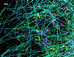 |
| This is a spectrally resolved super-resolution microscopy image of four subcellular targets that were labeled by four far-red dyes at 10 nm spectral separation. Color is used to indicate the measured fluorescence emission position of each single molecule. (Scale bar: 1 �m) CREDIT: Ke Xu/Berkeley Lab |
Abstract:
Using physical chemistry methods to look at biology at the nanoscale, a Lawrence Berkeley National Laboratory (Berkeley Lab) researcher has invented a new technology to image single molecules with unprecedented spectral and spatial resolution, thus leading to the first "true-color" super-resolution microscope.
Major innovation in molecular imaging delivers spatial and spectral info simultaneously: Berkeley Lab scientist invents technique to combine spectroscopy with super-resolution microscopy, enabling new ways to examine cell structures and study diseases
Berkeley, CA | Posted on August 17th, 2015Ke Xu, a faculty scientist in Berkeley Lab's Life Sciences Division, has dubbed his innovation SR-STORM, or spectrally resolved stochastic optical reconstruction microscopy. Because SR-STORM gives full spectral and spatial information for each molecule, the technology opens the door to high-resolution imaging of multiple components and local chemical environments, such as pH variations, inside a cell.
The research was reported in the journal Nature Methods in a paper titled, "Ultrahigh-throughput single-molecule spectroscopy and spectrally resolved super-resolution microscopy," with co-authors Zhengyang Zhang, Samuel Kenny, Margaret Hauser, and Wan Li, all of UC Berkeley. Xu is also an assistant professor at UC Berkeley's Department of Chemistry.
"We measure both the position and spectrum of each individual molecule, plotting its super-resolved spatial position in two dimensions and coloring each molecule according to its spectral position, so in that sense, it's true-color super-resolution microscopy, which is the first of its kind," Xu said. "This is a new type of imaging, combining single-molecule spectral measurement with super-resolution microscopy."
What's more, SR-STORM is high-throughput, able to deliver spatial and spectral information for millions of single molecules in about five minutes, compared to several minutes for a single frame of image comprising tens of molecules using conventional scanning-based techniques.
Xu built on work he did as a postdoctoral researcher at Harvard with Xiaowei Zhuang, who invented STORM, a super-resolution microscopy method based on single-molecule imaging and photoswitching. By devising a dual-objective system with two microscope lenses facing each other, Xu and colleagues viewed the front and back of the sample at the same time and achieved unprecedented optical resolution (of approximately 10 nanometers) of a cell. Using this method to image neurons, they showed that actin, a key component of the cytoskeleton (backbone of the cell), has a different structure in axons than in dendrites, two parts of a neuron.
But current super-resolution microscopy techniques do not deliver spectral information, which is useful for scientists to understand the behavior of individual molecules, as well as to enable high-quality multicolor imaging of multiple targets.
"So we constructed a dual-objective system but dispersed the single-molecule image collected by one objective lens into spectrum while keeping the other image for single-molecule localization," Xu said. "Now we are simultaneously accumulating the spectrum of the single molecules and also their position, so we solved the conundrum."
Next they dyed the sample with 14 different dyes in a narrow emission window and excited and photoswitched the molecules with one laser. While the spectra of the 14 dyes are heavily overlapping since they're close in emission, they found that the spectra of the individual molecules were surprisingly different and thus readily identifiable. "That's useful because it means we had a way to do multicolor imaging within a very narrow emission window," Xu said.
Indeed, using four dyes to label four different subcellular structures, such as mitochondria and microtubules, they were able to easily distinguish molecules of different dyes based on their spectral mean alone, and each subcellular structure was a distinct color.
"So using this method we can look at interactions between four biological components inside a cell in three-dimension and at very high resolution of about 10 nanometers," Xu said. "The applications are mostly in fundamental research and cell biology at this point, but hopefully it will lead to medical applications. This gives us new opportunities to look at cell structures, how they're built up, and whether there's any degradation of those structures in diseases."
Many diseases are caused either by an invading pathogen or degradation of a cell's internal structure. Alzheimer's, for example, may be related to degradation of the cytoskeleton inside neurons. "The cytoskeleton system is comprised of a host of interacting subcellular structures and proteins, and our technique will enable research on the interactions between these different targets with unprecedented number of color channels and spatial resolution," he said.
Next, Xu is trying to refine the method by using a single-objective system, and make it work with conventional microscope systems, thus making it more broadly accessible. He is also trying to develop suitable dyes and probes to monitor the local environment, such as the pH, in live cells at the nanometer scale.
###
The research was partly supported by UC Berkeley's College of Chemistry and a Laboratory Directed Research and Development (LDRD) grant by Berkeley Lab.
####
About DOE/Lawrence Berkeley National Laboratory
Lawrence Berkeley National Laboratory addresses the world's most urgent scientific challenges by advancing sustainable energy, protecting human health, creating new materials, and revealing the origin and fate of the universe. Founded in 1931, Berkeley Lab's scientific expertise has been recognized with 13 Nobel prizes. The University of California manages Berkeley Lab for the U.S. Department of Energy's Office of Science. For more, visit www.lbl.gov.
DOE's Office of Science is the single largest supporter of basic research in the physical sciences in the United States, and is working to address some of the most pressing challenges of our time. For more information, please visit science.energy.gov.
For more information, please click here
Contacts:
Julie Chao
510-486-6491
Copyright © DOE/Lawrence Berkeley National Laboratory
If you have a comment, please Contact us.Issuers of news releases, not 7th Wave, Inc. or Nanotechnology Now, are solely responsible for the accuracy of the content.
Bookmark:


| Related News Press |
News and information
![]() Quantum computer improves AI predictions April 17th, 2026
Quantum computer improves AI predictions April 17th, 2026
![]() Flexible sensor gains sensitivity under pressure April 17th, 2026
Flexible sensor gains sensitivity under pressure April 17th, 2026
![]() A reusable chip for particulate matter sensing April 17th, 2026
A reusable chip for particulate matter sensing April 17th, 2026
![]() Detecting vibrational quantum beating in the predissociation dynamics of SF6 using time-resolved photoelectron spectroscopy April 17th, 2026
Detecting vibrational quantum beating in the predissociation dynamics of SF6 using time-resolved photoelectron spectroscopy April 17th, 2026
Imaging
![]() Simple algorithm paired with standard imaging tool could predict failure in lithium metal batteries August 8th, 2025
Simple algorithm paired with standard imaging tool could predict failure in lithium metal batteries August 8th, 2025
![]() First real-time observation of two-dimensional melting process: Researchers at Mainz University unveil new insights into magnetic vortex structures August 8th, 2025
First real-time observation of two-dimensional melting process: Researchers at Mainz University unveil new insights into magnetic vortex structures August 8th, 2025
![]() New imaging approach transforms study of bacterial biofilms August 8th, 2025
New imaging approach transforms study of bacterial biofilms August 8th, 2025
Laboratories
![]() Researchers develop molecular qubits that communicate at telecom frequencies October 3rd, 2025
Researchers develop molecular qubits that communicate at telecom frequencies October 3rd, 2025
Govt.-Legislation/Regulation/Funding/Policy
![]() Quantum computer improves AI predictions April 17th, 2026
Quantum computer improves AI predictions April 17th, 2026
![]() Metasurfaces smooth light to boost magnetic sensing precision January 30th, 2026
Metasurfaces smooth light to boost magnetic sensing precision January 30th, 2026
![]() New imaging approach transforms study of bacterial biofilms August 8th, 2025
New imaging approach transforms study of bacterial biofilms August 8th, 2025
Discoveries
![]() Quantum computer improves AI predictions April 17th, 2026
Quantum computer improves AI predictions April 17th, 2026
![]() Flexible sensor gains sensitivity under pressure April 17th, 2026
Flexible sensor gains sensitivity under pressure April 17th, 2026
![]() A reusable chip for particulate matter sensing April 17th, 2026
A reusable chip for particulate matter sensing April 17th, 2026
![]() Detecting vibrational quantum beating in the predissociation dynamics of SF6 using time-resolved photoelectron spectroscopy April 17th, 2026
Detecting vibrational quantum beating in the predissociation dynamics of SF6 using time-resolved photoelectron spectroscopy April 17th, 2026
Announcements
![]() A fundamentally new therapeutic approach to cystic fibrosis: Nanobody repairs cellular defect April 17th, 2026
A fundamentally new therapeutic approach to cystic fibrosis: Nanobody repairs cellular defect April 17th, 2026
![]() UC Irvine physicists discover method to reverse �quantum scrambling� : The work addresses the problem of information loss in quantum computing system April 17th, 2026
UC Irvine physicists discover method to reverse �quantum scrambling� : The work addresses the problem of information loss in quantum computing system April 17th, 2026
Tools
![]() Metasurfaces smooth light to boost magnetic sensing precision January 30th, 2026
Metasurfaces smooth light to boost magnetic sensing precision January 30th, 2026
![]() From sensors to smart systems: the rise of AI-driven photonic noses January 30th, 2026
From sensors to smart systems: the rise of AI-driven photonic noses January 30th, 2026
![]() Japan launches fully domestically produced quantum computer: Expo visitors to experience quantum computing firsthand August 8th, 2025
Japan launches fully domestically produced quantum computer: Expo visitors to experience quantum computing firsthand August 8th, 2025
Research partnerships
![]() Lab to industry: InSe wafer-scale breakthrough for future electronics August 8th, 2025
Lab to industry: InSe wafer-scale breakthrough for future electronics August 8th, 2025
![]() HKU physicists uncover hidden order in the quantum world through deconfined quantum critical points April 25th, 2025
HKU physicists uncover hidden order in the quantum world through deconfined quantum critical points April 25th, 2025
|
|
||
|
|
||
| The latest news from around the world, FREE | ||
|
|
||
|
|
||
| Premium Products | ||
|
|
||
|
Only the news you want to read!
Learn More |
||
|
|
||
|
Full-service, expert consulting
Learn More |
||
|
|
||