Home > Press > Membrane nano-tomography in living cells: Label-free evanescent microscopy enables full-field and real-time tracking of membrane processes without signal fading and cell perturbation
 |
Abstract:
Membranes play a pivotal role in numerous cell mechanisms, in particular for internalization, adhesion and motility studies. In terms of optical imaging of the membrane, special configurations are needed to remove the light coming from the inner part of the cell. French scientists now show that through-the-objective evanescent microscopy (epi-EM) is a powerful technique to image membranes in living cells.
Membrane nano-tomography in living cells: Label-free evanescent microscopy enables full-field and real-time tracking of membrane processes without signal fading and cell perturbation
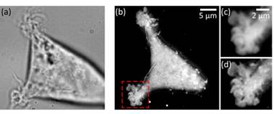
Paris, France | Posted on December 5th, 2014In label-free evanescent microscopy (EM), configurations similar to total internal reflection fluorescence (TIRF) have been proposed: prism-based or through-the-objective. However, in the latter case, these evanescent techniques have not spread much, with a relative preference for the prism-based configuration also called total internal reflection microscopy (TIRM). The team led by Pierre Bon chose the through-the-objective based configuration (epi-EM), which enabled super-axially resolved tomographic reconstruction of the basal membrane of label-free living cells. The implementation of epi-EM only required an easy to settle illumination/collection scheme on a standard inverted microscope. Only a high-NA objective (NAobj > 1.33) was needed for living biological sample studies and a spatial filter on the epi-illumination arm in order to reject under-critical angle illumination.
Either bead calibration or a multilayer Fresnel model could be used to retrieve nanometric position. Based on a multilayer Fresnel model, the team was able to retrieve the membrane/interface distance with 10 nm precision. The researchers applied this nano-axial tomography to retrieve quantitative information on invagination dynamics of living cell membranes. They studied the membrane elevation map of living cells (Wt HEK-293) during 15 minutes at one frame per second without perturbing the sample.
The results demonstrate that epi-EM gives easily access to axially super-resolved images of unlabeled microscopic samples with almost no microscope modification, and at least a doubled lateral resolution compared to classical TIRM. A study can be of any duration as the signal level is not sensitive to any fluorophore stability dependence and the photoxicity is very low as barely any light is absorbed by the sample. The scientists are convinced that this technique will be useful for cell motility and adhesion studies when the sample cannot be modified (ex. stem cells) or when very fast and/or long studies are required.
(Text contributed by K. Maedefessel-Herrmann)
####
About Journal of Biophotonics
Journal of Biophotonics publishes cutting edge research on interactions between light and biological material. The journal is highly interdisciplinary, covering research in the fields of physics, chemistry, biology and medicine. The scope extends from basic research to clinical applications. Connecting scientists who try to understand basic biological processes using light as a diagnostic and therapeutic tool, the journal offers a platform where the physicist communicates with the biologist and where the clinical practitioner learns about the latest tools for diagnosis of diseases. JBP offers fast publication times: down to 20 days from acceptance to publication. Impact Factor 2013: 3.856
For more information, please click here
Contacts:
Regina Hagen
Journal Publishing Manager
JBP
Copyright © Journal of Biophotonics
If you have a comment, please Contact us.Issuers of news releases, not 7th Wave, Inc. or Nanotechnology Now, are solely responsible for the accuracy of the content.
Bookmark:


| Related Links |
| Related News Press |
News and information
![]() Quantum computer improves AI predictions April 17th, 2026
Quantum computer improves AI predictions April 17th, 2026
![]() Flexible sensor gains sensitivity under pressure April 17th, 2026
Flexible sensor gains sensitivity under pressure April 17th, 2026
![]() A reusable chip for particulate matter sensing April 17th, 2026
A reusable chip for particulate matter sensing April 17th, 2026
![]() Detecting vibrational quantum beating in the predissociation dynamics of SF6 using time-resolved photoelectron spectroscopy April 17th, 2026
Detecting vibrational quantum beating in the predissociation dynamics of SF6 using time-resolved photoelectron spectroscopy April 17th, 2026
Imaging
![]() Simple algorithm paired with standard imaging tool could predict failure in lithium metal batteries August 8th, 2025
Simple algorithm paired with standard imaging tool could predict failure in lithium metal batteries August 8th, 2025
Nanomedicine
![]() A fundamentally new therapeutic approach to cystic fibrosis: Nanobody repairs cellular defect April 17th, 2026
A fundamentally new therapeutic approach to cystic fibrosis: Nanobody repairs cellular defect April 17th, 2026
![]() New molecular technology targets tumors and simultaneously silences two �undruggable� cancer genes August 8th, 2025
New molecular technology targets tumors and simultaneously silences two �undruggable� cancer genes August 8th, 2025
![]() New imaging approach transforms study of bacterial biofilms August 8th, 2025
New imaging approach transforms study of bacterial biofilms August 8th, 2025
![]() Electrifying results shed light on graphene foam as a potential material for lab grown cartilage June 6th, 2025
Electrifying results shed light on graphene foam as a potential material for lab grown cartilage June 6th, 2025
Discoveries
![]() Quantum computer improves AI predictions April 17th, 2026
Quantum computer improves AI predictions April 17th, 2026
![]() Flexible sensor gains sensitivity under pressure April 17th, 2026
Flexible sensor gains sensitivity under pressure April 17th, 2026
![]() A reusable chip for particulate matter sensing April 17th, 2026
A reusable chip for particulate matter sensing April 17th, 2026
![]() Detecting vibrational quantum beating in the predissociation dynamics of SF6 using time-resolved photoelectron spectroscopy April 17th, 2026
Detecting vibrational quantum beating in the predissociation dynamics of SF6 using time-resolved photoelectron spectroscopy April 17th, 2026
Announcements
![]() A fundamentally new therapeutic approach to cystic fibrosis: Nanobody repairs cellular defect April 17th, 2026
A fundamentally new therapeutic approach to cystic fibrosis: Nanobody repairs cellular defect April 17th, 2026
![]() UC Irvine physicists discover method to reverse �quantum scrambling� : The work addresses the problem of information loss in quantum computing system April 17th, 2026
UC Irvine physicists discover method to reverse �quantum scrambling� : The work addresses the problem of information loss in quantum computing system April 17th, 2026
Interviews/Book Reviews/Essays/Reports/Podcasts/Journals/White papers/Posters
![]() A fundamentally new therapeutic approach to cystic fibrosis: Nanobody repairs cellular defect April 17th, 2026
A fundamentally new therapeutic approach to cystic fibrosis: Nanobody repairs cellular defect April 17th, 2026
![]() UC Irvine physicists discover method to reverse �quantum scrambling� : The work addresses the problem of information loss in quantum computing system April 17th, 2026
UC Irvine physicists discover method to reverse �quantum scrambling� : The work addresses the problem of information loss in quantum computing system April 17th, 2026
Tools
![]() Metasurfaces smooth light to boost magnetic sensing precision January 30th, 2026
Metasurfaces smooth light to boost magnetic sensing precision January 30th, 2026
![]() From sensors to smart systems: the rise of AI-driven photonic noses January 30th, 2026
From sensors to smart systems: the rise of AI-driven photonic noses January 30th, 2026
![]() Japan launches fully domestically produced quantum computer: Expo visitors to experience quantum computing firsthand August 8th, 2025
Japan launches fully domestically produced quantum computer: Expo visitors to experience quantum computing firsthand August 8th, 2025
|
|
||
|
|
||
| The latest news from around the world, FREE | ||
|
|
||
|
|
||
| Premium Products | ||
|
|
||
|
Only the news you want to read!
Learn More |
||
|
|
||
|
Full-service, expert consulting
Learn More |
||
|
|
||