Home > Press > Nanoscale freezing leads to better imaging
 |
| Images of a frozen-hydrated algae cell. (a) Some cell ultrastructure is shown using differential phase contrast imaging. (b) Distributions of zinc, iron, and potassium are visible in this X-ray fluorescence image. |
Abstract:
It's an odd twist. For scientists to determine if a cell is functioning properly, they must destroy it.
Nanoscale freezing leads to better imaging
Argonne, IL | Posted on February 26th, 2014This is what happens in X-ray fluorescence microscopy when biological specimens are exposed to ionizing radiation, which provides images with a level of detail that conventional microscopes just can't match. This exposure can change what is being imaged in profound ways, possibly giving false accounts of how the cell actually works.
To address this issue, researchers at the U.S. Department of Energy's (DOE) Argonne National Laboratory created a new probe that freezes cells to "see" at greater detail without damaging the sample.
The issue boils down to preparation.
Traditional X-ray methods look at cells that have either been immersed in water or dehydrated, like astronaut food. For wet specimens at room temperature, the radiation can break the bonds linking molecules together and cause them to scatter, changing the sample's structure.
For dehydrated specimens, potassium and other diffusible ions are washed away during chemical fixation, which kills the cell and loosens the cell membrane, allowing ions to escape. Moreover, when the sample is dehydrated, the cell can shrink, distort or even collapse.
"Imagine a ball. When you dry it, you make it flat," says Si Chen, principle author of the study. "It changes the structure of the sample and also the distribution of the trace elements that we are looking for."
To address this issue, Argonne researchers developed a hard X-ray fluorescence nanoprobe called the Bionanoprobe, which makes three-dimensional images that map out the locations of trace elements, like iron or potassium, in frozen biological samples.
"We don't want to dry the sample; we want to keep it hydrated," says Chen. "We plunge the sample into liquid ethane at very high speeds and then look at the frozen sample directly."
Rapidly cooling biological specimens to temperatures of -260�F preserves the natural state of a cell's organelles and trace elements while retaining the water in the sample.
Housed at an undulator beamline at sector 21 of Argonne's Advanced Photon Source, the Bionanoprobe features a vacuum chamber that eliminates frosting and convective heating and automatically acquires tomographic (sectioned images) data sets. Sector 21 is sponsored by a consortium of several universities and a research institute known collectively as the Life Sciences Collaborative Action Team.
The Bionanoprobe can also produce extremely high-resolution images at the smallest scales�below 100 nanometers. Compare that to a typical human hair, which is 80,000 to 100,000 nanometers wide. Chen uses X-ray optics called zone plates to focus the X-ray beam down to a miniscule small spot. A simple scan produces an image with a full fluorescent spectrum for each scanning step.
Recent tests have been encouraging. One team of researchers successfully acquired differential phase contrast and X-ray fluorescence images simultaneously by raster scanning of a green algae. The former gave researchers some of the algae's ultrastructure, and using the latter, they were able to show evenly distributed potassium and patterned distributions of zinc and iron.
"We can see the trace element distribution, but with biological samples, the contrast from the structure is typically very low," says Chen. "Phase contrast imaging highlights the structural details."
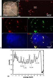
Another study made X-ray fluorescence images of an immortal cervical cancer cell line called HeLa cells. The samples were plunge-frozen, chemically fixed and then treated with an iron oxide core in a titanium dioxide shell nanocomposite, which allowed researchers to determine if the nanocomposites actually made it into the cell nucleus.
Dr. Gale Woloschak, professor at Northwestern University's Feinberg School of Medicine conducted the study. She created nanoparticles that target and kill cancer cells, but when the researchers wanted to see where the nanoparticles actually wound up in the cell, they ran into trouble with traditional X-ray methods.
"This is the problem," says Woloschak. "If you think of how two-dimensional X-ray imaging works, X-rays penetrate through the entire cell, so it's hard to determine whether the nanoparticles are above, below or inside the nucleus. What the Bionanoprobe does is give us a three-dimensional image�we could actually see that the nanoparticles were imbedded in the nucleus."
The work is reported in "The Bionanoprobe: hard X-ray fluorescence nanoprobe with cryogenic capabilities," published last month in the Journal of Synchrotron Radiation.
This work was supported with funding by the National Institutes of Health. Use of the Advanced Photon Source is funded by the Department of Energy's Office of Science.
####
About DOE/Argonne National Laboratory
Argonne National Laboratory seeks solutions to pressing national problems in science and technology. The nation's first national laboratory, Argonne conducts leading-edge basic and applied scientific research in virtually every scientific discipline. Argonne researchers work closely with researchers from hundreds of companies, universities, and federal, state and municipal agencies to help them solve their specific problems, advance America's scientific leadership and prepare the nation for a better future. With employees from more than 60 nations, Argonne is managed by UChicago Argonne, LLC for the U.S. Department of Energy's Office of Science.
The Advanced Photon Source at Argonne National Laboratory is one of five national synchrotron radiation light sources supported by the U.S. Department of Energy�s Office of Science to carry out applied and basic research to understand, predict, and ultimately control matter and energy at the electronic, atomic, and molecular levels, provide the foundations for new energy technologies, and support DOE missions in energy, environment, and national security. To learn more about the Office of Science X-ray user facilities, visit the user facilities directory.
The Life Sciences Collaborative Access Team (LS-CAT) provides macromolecular crystallography resources at Argonne National Laboratory�s Advanced Photon Source for those with a need to determine the structure of proteins. Current LS-CAT members are Michigan State University, University of Michigan, Wayne State University, Van Andel Research Institute, Northwestern University, University Wisconsin-Madison, Vanderbilt University, and University of Illinois at Urbana-Champaign.
DOE�s Office of Science is the single largest supporter of basic research in the physical sciences in the United States, and is working to address some of the most pressing challenges of our time. For more information, please visit science.energy.gov.
For more information, please click here
Contacts:
Tona Kunz
630-252-5560
Jared Sagoff
(630) 252-5549
Copyright © DOE/Argonne National Laboratory
If you have a comment, please Contact us.Issuers of news releases, not 7th Wave, Inc. or Nanotechnology Now, are solely responsible for the accuracy of the content.
Bookmark:


| Related News Press |
News and information
![]() Quantum computer improves AI predictions April 17th, 2026
Quantum computer improves AI predictions April 17th, 2026
![]() Flexible sensor gains sensitivity under pressure April 17th, 2026
Flexible sensor gains sensitivity under pressure April 17th, 2026
![]() A reusable chip for particulate matter sensing April 17th, 2026
A reusable chip for particulate matter sensing April 17th, 2026
![]() Detecting vibrational quantum beating in the predissociation dynamics of SF6 using time-resolved photoelectron spectroscopy April 17th, 2026
Detecting vibrational quantum beating in the predissociation dynamics of SF6 using time-resolved photoelectron spectroscopy April 17th, 2026
Imaging
![]() Simple algorithm paired with standard imaging tool could predict failure in lithium metal batteries August 8th, 2025
Simple algorithm paired with standard imaging tool could predict failure in lithium metal batteries August 8th, 2025
![]() First real-time observation of two-dimensional melting process: Researchers at Mainz University unveil new insights into magnetic vortex structures August 8th, 2025
First real-time observation of two-dimensional melting process: Researchers at Mainz University unveil new insights into magnetic vortex structures August 8th, 2025
![]() New imaging approach transforms study of bacterial biofilms August 8th, 2025
New imaging approach transforms study of bacterial biofilms August 8th, 2025
Laboratories
![]() Researchers develop molecular qubits that communicate at telecom frequencies October 3rd, 2025
Researchers develop molecular qubits that communicate at telecom frequencies October 3rd, 2025
Govt.-Legislation/Regulation/Funding/Policy
![]() Quantum computer improves AI predictions April 17th, 2026
Quantum computer improves AI predictions April 17th, 2026
![]() Metasurfaces smooth light to boost magnetic sensing precision January 30th, 2026
Metasurfaces smooth light to boost magnetic sensing precision January 30th, 2026
![]() New imaging approach transforms study of bacterial biofilms August 8th, 2025
New imaging approach transforms study of bacterial biofilms August 8th, 2025
Discoveries
![]() Quantum computer improves AI predictions April 17th, 2026
Quantum computer improves AI predictions April 17th, 2026
![]() Flexible sensor gains sensitivity under pressure April 17th, 2026
Flexible sensor gains sensitivity under pressure April 17th, 2026
![]() A reusable chip for particulate matter sensing April 17th, 2026
A reusable chip for particulate matter sensing April 17th, 2026
![]() Detecting vibrational quantum beating in the predissociation dynamics of SF6 using time-resolved photoelectron spectroscopy April 17th, 2026
Detecting vibrational quantum beating in the predissociation dynamics of SF6 using time-resolved photoelectron spectroscopy April 17th, 2026
Announcements
![]() A fundamentally new therapeutic approach to cystic fibrosis: Nanobody repairs cellular defect April 17th, 2026
A fundamentally new therapeutic approach to cystic fibrosis: Nanobody repairs cellular defect April 17th, 2026
![]() UC Irvine physicists discover method to reverse �quantum scrambling� : The work addresses the problem of information loss in quantum computing system April 17th, 2026
UC Irvine physicists discover method to reverse �quantum scrambling� : The work addresses the problem of information loss in quantum computing system April 17th, 2026
Tools
![]() Metasurfaces smooth light to boost magnetic sensing precision January 30th, 2026
Metasurfaces smooth light to boost magnetic sensing precision January 30th, 2026
![]() From sensors to smart systems: the rise of AI-driven photonic noses January 30th, 2026
From sensors to smart systems: the rise of AI-driven photonic noses January 30th, 2026
![]() Japan launches fully domestically produced quantum computer: Expo visitors to experience quantum computing firsthand August 8th, 2025
Japan launches fully domestically produced quantum computer: Expo visitors to experience quantum computing firsthand August 8th, 2025
|
|
||
|
|
||
| The latest news from around the world, FREE | ||
|
|
||
|
|
||
| Premium Products | ||
|
|
||
|
Only the news you want to read!
Learn More |
||
|
|
||
|
Full-service, expert consulting
Learn More |
||
|
|
||