Home > Press > Bruker Introduces High-Performance Opterra™ Multipoint Scanning Confocal Microscope: Opterra Offers Superior Integration of Confocal Microscopy and Photoactivation for Biology Applications
 |
Abstract:
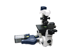
Today at the 2013 American Society for Cell Biology Annual Meeting, Bruker introduced the Opterra Multipoint Scanning Confocal Microscope, which sets a new standard for integration of confocal imaging with photoactivation. The new Opterra microscope utilizes a number of innovative features to obtain the speed of wide-field imaging and the resolution of traditional confocal systems while minimizing phototoxicity, making it an ideal solution for gentle and fast confocal imaging of live cell preparations. A seven-position pinhole/slit aperture allows the Opterra to be optimized for varying objective lens magnifications that results in the ability to image deeper into tissue versus conventional disk scanning confocal microscopes.
Bruker Introduces High-Performance Opterra™ Multipoint Scanning Confocal Microscope: Opterra Offers Superior Integration of Confocal Microscopy and Photoactivation for Biology Applications
New Orleans, LA | Posted on December 16th, 2013"The Opterra has proven to be a major advance in terms of rapid, time-based volumetric imaging," said Dr. Mario De Bono, Medical Research Council Group Leader at the Laboratory of Molecular Biology, Cambridge University, UK. "The speed of the system, coupled with its sensitivity and resolution has significantly enhanced our ability to visualize neural activity in 3D in C. elegans at speeds that were previously not possible. The ability to change pinhole size is great, as it allows us to match the imaging setup with the specimen."
"Our new Opterra provides a flexible optical workstation for cell biologists to perform confocal imaging of live cells and small organisms with simultaneous point and area scanning for photoactivation and photoablation," explained Mike Szulczewski, Vice President and General Manager of Bruker's Fluorescence Microscopy business. "The tight integration of optical imaging with optical stimulation techniques enables investigators to take full advantage of today's imaging and photochemical probe technologies."
About Opterra
The Opterra Multipoint Scanning Confocal Microscope is based on Bruker's patented swept-field imaging scanner. This scanner allows high-speed confocal imaging of live cell and small organism preparations at resolutions comparable to conventional point scanners, but with minimal phototoxicty. Opterra includes a second scanner for photo- activation/bleaching/ablation, which can operate simultaneously with imaging. The photoactivation scanner can be coupled to both visible and multiphoton lasers, thus allowing the use of the full range of photo-activatable molecules and photochemical techniques available to life science researchers. In the case of multiphoton lasers, this provides precise three-dimensional control over photoactivation. The applications addressed by Opterra include response to DNA damage, kinetics of photoactivatable fluorescent proteins, fluorescence recovery after photo-bleaching (FRAP), response to local stimulation of channel proteins, and response to cell membrane damage. Bruker's Prairie View 5.0 software provides an intuitive interface with a rich environment for defining image acquisition and photoactivation protocols.
####
About Bruker Corporation
Bruker Corporation is a leading provider of high-performance scientific instruments and solutions for molecular and materials research, as well as for industrial and applied analysis.
For more information, please click here
Contacts:
Stephen Hopkins, Marketing Communications
Bruker Nano Surfaces Division
3400 East Britannia Drive, Suite 150, Tucson, AZ 85706
T: +1 (520) 741-1044 x1022
Copyright © Bruker Corporation
If you have a comment, please Contact us.Issuers of news releases, not 7th Wave, Inc. or Nanotechnology Now, are solely responsible for the accuracy of the content.
Bookmark:


| Related News Press |
News and information
![]() Quantum computer improves AI predictions April 17th, 2026
Quantum computer improves AI predictions April 17th, 2026
![]() Flexible sensor gains sensitivity under pressure April 17th, 2026
Flexible sensor gains sensitivity under pressure April 17th, 2026
![]() A reusable chip for particulate matter sensing April 17th, 2026
A reusable chip for particulate matter sensing April 17th, 2026
![]() Detecting vibrational quantum beating in the predissociation dynamics of SF6 using time-resolved photoelectron spectroscopy April 17th, 2026
Detecting vibrational quantum beating in the predissociation dynamics of SF6 using time-resolved photoelectron spectroscopy April 17th, 2026
Nanomedicine
![]() A fundamentally new therapeutic approach to cystic fibrosis: Nanobody repairs cellular defect April 17th, 2026
A fundamentally new therapeutic approach to cystic fibrosis: Nanobody repairs cellular defect April 17th, 2026
![]() New molecular technology targets tumors and simultaneously silences two ‘undruggable’ cancer genes August 8th, 2025
New molecular technology targets tumors and simultaneously silences two ‘undruggable’ cancer genes August 8th, 2025
![]() New imaging approach transforms study of bacterial biofilms August 8th, 2025
New imaging approach transforms study of bacterial biofilms August 8th, 2025
![]() Electrifying results shed light on graphene foam as a potential material for lab grown cartilage June 6th, 2025
Electrifying results shed light on graphene foam as a potential material for lab grown cartilage June 6th, 2025
Announcements
![]() A fundamentally new therapeutic approach to cystic fibrosis: Nanobody repairs cellular defect April 17th, 2026
A fundamentally new therapeutic approach to cystic fibrosis: Nanobody repairs cellular defect April 17th, 2026
![]() UC Irvine physicists discover method to reverse ‘quantum scrambling’ : The work addresses the problem of information loss in quantum computing system April 17th, 2026
UC Irvine physicists discover method to reverse ‘quantum scrambling’ : The work addresses the problem of information loss in quantum computing system April 17th, 2026
Tools
![]() Metasurfaces smooth light to boost magnetic sensing precision January 30th, 2026
Metasurfaces smooth light to boost magnetic sensing precision January 30th, 2026
![]() From sensors to smart systems: the rise of AI-driven photonic noses January 30th, 2026
From sensors to smart systems: the rise of AI-driven photonic noses January 30th, 2026
![]() Japan launches fully domestically produced quantum computer: Expo visitors to experience quantum computing firsthand August 8th, 2025
Japan launches fully domestically produced quantum computer: Expo visitors to experience quantum computing firsthand August 8th, 2025
Events/Classes
![]() Institute for Nanoscience hosts annual proposal planning meeting May 16th, 2025
Institute for Nanoscience hosts annual proposal planning meeting May 16th, 2025
![]() A New Blue: Mysterious origin of the ribbontail ray’s electric blue spots revealed July 5th, 2024
A New Blue: Mysterious origin of the ribbontail ray’s electric blue spots revealed July 5th, 2024
![]() Researchers demonstrate co-propagation of quantum and classical signals: Study shows that quantum encryption can be implemented in existing fiber networks January 20th, 2023
Researchers demonstrate co-propagation of quantum and classical signals: Study shows that quantum encryption can be implemented in existing fiber networks January 20th, 2023
|
|
||
|
|
||
| The latest news from around the world, FREE | ||
|
|
||
|
|
||
| Premium Products | ||
|
|
||
|
Only the news you want to read!
Learn More |
||
|
|
||
|
Full-service, expert consulting
Learn More |
||
|
|
||