Home > Press > So BRIGHT, you need to wear shades: Tiny probes shine brightly to reveal the location of targeted tissues
 |
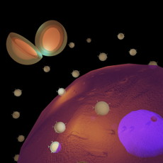
| NAVEEN GANDRA Nanostructures called BRIGHTs seek out biomarkers on cells and then beam brightly to reveal their locations. In the tiny gap between the gold skin and the gold core of the cleaved BRIGHT (visible to the upper left), there is an electromagnetic hot spot that lights up the reporter molecules trapped there. |
Abstract:
Called BRIGHTs, the tiny probes described in the online issue of Advanced Materials on Nov. 15, bind to biomarkers of disease and, when swept by an infrared laser, light up to reveal their location.
So BRIGHT, you need to wear shades: Tiny probes shine brightly to reveal the location of targeted tissues
St. Louis, MO | Posted on November 20th, 2012Tiny as they are, the probes are exquisitely engineered objects: gold nanoparticles covered with molecules called Raman reporters, in turn covered by a thin shell of gold that spontaneously forms a dodecahedron.
The Raman reporters are molecules whose jiggling atoms respond to a probe laser by scattering light at characteristic wavelengths.
The shell and core create an electromagnetic hotspot in the gap between them that boosts the reporters' emission by a factor of nearly a trillion.
BRIGHTs shine about 1.7 x 10¹¹ more brightly than isolated Raman reporters and about 20 times more intensely than the next-closest competitor probe, says Srikanth Singamaneni, PhD, assistant professor of mechanical engineering and materials science in the School of Engineering & Applied Science at Washington University in St. Louis.
Goosing the signal from Raman reporters
Singamaneni and his postdoctoral research associate Naveen Gandra, PhD, tried several different probe designs before settling on BRIGHTS.
Singamaneni's lab has worked for years with Raman spectroscopy, a spectroscopic technique that is used to study the vibrational modes (bending and stretching) of molecules. Laser light interacts with these modes and the molecule then emits light at higher or lower wavelengths that are characteristic of the molecule,
Spontaneous Raman scattering, as this phenomenon is called, is by nature very weak, but 30 years ago scientists accidently stumbled on the fact that it is much stronger if the molecules are adsorbed on roughened metallic surfaces. Then they discovered that molecules attached to metallic nanoparticles shine even brighter than those attached to rough surfaces.
The intensity boost from surface-enhanced Raman scattering, or SERS, is potentially huge. "It's well-known that if you sandwich Raman reporters between two plasmonic materials, such as gold or silver, you are going to see dramatic Raman enhancement," Singamaneni says.
Originally his team tried to create intense electromagnetic hot spots by sticking smaller particles onto a larger central particle, creating core-satellite assemblies that look like daisies.
"But we realized these assemblies are not ideal for bioimaging," he says, "because the particles were held together by weak electrostatic interactions and the assemblies were going to come apart in the body."
Next they tried using something called Click chemistry to make stronger covalent bonds between the satellites and the core.
"We had some success with those assemblies," Singamaneni says, "but in the meantime we had started to wonder if we couldn't make an electromagnetic hot spot within a single nanoparticle rather than among particles.
"It occurred to us that if we put Raman reporters between the core and shell of a single particle could we create an internal hotspot."
That idea worked like a charm.
A rainbow of probes carefully dispensing drugs?
The next step, says Singamaneni, is to test BRIGHTS in vivo in the lab of Sam Achilefu, PhD, professor of radiology in the School of Medicine.
But he's already thinking of ways to get even more out of the design.
Since different Raman reporter molecules respond at different wavelengths, Singamaneni says, it should be possible to design BRIGHTS targeted to different biomolecules that also have different Raman reporters and then monitor them all simultaneously with the same light probe.
And he and Gandra would like to combine BRIGHTS with a drug container of some kind, so that the containers could be tracked in the body and the drug and released only when it reached the target tissue, thus avoiding many of the side effects patients dread.
Good things, as they say, come in small packages.
####
For more information, please click here
Contacts:
Diana Lutz
314-935-5272
Copyright © Washington University in St. Louis
If you have a comment, please Contact us.Issuers of news releases, not 7th Wave, Inc. or Nanotechnology Now, are solely responsible for the accuracy of the content.
| Related News Press |
News and information
![]() Researchers develop molecular qubits that communicate at telecom frequencies October 3rd, 2025
Researchers develop molecular qubits that communicate at telecom frequencies October 3rd, 2025
![]() Next-generation quantum communication October 3rd, 2025
Next-generation quantum communication October 3rd, 2025
![]() "Nanoreactor" cage uses visible light for catalytic and ultra-selective cross-cycloadditions October 3rd, 2025
"Nanoreactor" cage uses visible light for catalytic and ultra-selective cross-cycloadditions October 3rd, 2025
Imaging
![]() ICFO researchers overcome long-standing bottleneck in single photon detection with twisted 2D materials August 8th, 2025
ICFO researchers overcome long-standing bottleneck in single photon detection with twisted 2D materials August 8th, 2025
![]() Simple algorithm paired with standard imaging tool could predict failure in lithium metal batteries August 8th, 2025
Simple algorithm paired with standard imaging tool could predict failure in lithium metal batteries August 8th, 2025
![]() First real-time observation of two-dimensional melting process: Researchers at Mainz University unveil new insights into magnetic vortex structures August 8th, 2025
First real-time observation of two-dimensional melting process: Researchers at Mainz University unveil new insights into magnetic vortex structures August 8th, 2025
![]() New imaging approach transforms study of bacterial biofilms August 8th, 2025
New imaging approach transforms study of bacterial biofilms August 8th, 2025
Nanomedicine
![]() New molecular technology targets tumors and simultaneously silences two ‘undruggable’ cancer genes August 8th, 2025
New molecular technology targets tumors and simultaneously silences two ‘undruggable’ cancer genes August 8th, 2025
![]() New imaging approach transforms study of bacterial biofilms August 8th, 2025
New imaging approach transforms study of bacterial biofilms August 8th, 2025
![]() Cambridge chemists discover simple way to build bigger molecules – one carbon at a time June 6th, 2025
Cambridge chemists discover simple way to build bigger molecules – one carbon at a time June 6th, 2025
![]() Electrifying results shed light on graphene foam as a potential material for lab grown cartilage June 6th, 2025
Electrifying results shed light on graphene foam as a potential material for lab grown cartilage June 6th, 2025
Discoveries
![]() Researchers develop molecular qubits that communicate at telecom frequencies October 3rd, 2025
Researchers develop molecular qubits that communicate at telecom frequencies October 3rd, 2025
![]() Next-generation quantum communication October 3rd, 2025
Next-generation quantum communication October 3rd, 2025
![]() "Nanoreactor" cage uses visible light for catalytic and ultra-selective cross-cycloadditions October 3rd, 2025
"Nanoreactor" cage uses visible light for catalytic and ultra-selective cross-cycloadditions October 3rd, 2025
Announcements
![]() Rice membrane extracts lithium from brines with greater speed, less waste October 3rd, 2025
Rice membrane extracts lithium from brines with greater speed, less waste October 3rd, 2025
![]() Researchers develop molecular qubits that communicate at telecom frequencies October 3rd, 2025
Researchers develop molecular qubits that communicate at telecom frequencies October 3rd, 2025
![]() Next-generation quantum communication October 3rd, 2025
Next-generation quantum communication October 3rd, 2025
![]() "Nanoreactor" cage uses visible light for catalytic and ultra-selective cross-cycloadditions October 3rd, 2025
"Nanoreactor" cage uses visible light for catalytic and ultra-selective cross-cycloadditions October 3rd, 2025
Tools
![]() Japan launches fully domestically produced quantum computer: Expo visitors to experience quantum computing firsthand August 8th, 2025
Japan launches fully domestically produced quantum computer: Expo visitors to experience quantum computing firsthand August 8th, 2025
![]() Rice researchers harness gravity to create low-cost device for rapid cell analysis February 28th, 2025
Rice researchers harness gravity to create low-cost device for rapid cell analysis February 28th, 2025
Photonics/Optics/Lasers
![]() ICFO researchers overcome long-standing bottleneck in single photon detection with twisted 2D materials August 8th, 2025
ICFO researchers overcome long-standing bottleneck in single photon detection with twisted 2D materials August 8th, 2025
![]() Institute for Nanoscience hosts annual proposal planning meeting May 16th, 2025
Institute for Nanoscience hosts annual proposal planning meeting May 16th, 2025
|
|
||
|
|
||
| The latest news from around the world, FREE | ||
|
|
||
|
|
||
| Premium Products | ||
|
|
||
|
Only the news you want to read!
Learn More |
||
|
|
||
|
Full-service, expert consulting
Learn More |
||
|
|
||


