Home > Press > FEI Introduces New Correlative Workflow Solution for Life Sciences: MAPS software combines the advantages of both light and electron microscopy for cellular biology research.
 |
Abstract:
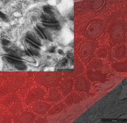
FEI (NASDAQ: FEIC), a leading instrumentation company providing imaging and analysis systems for research and industry, today announced a new correlative workflow solution for research scientists. FEI's MAPS (Modular Automated Processing System) provides a fast and efficient correlative workflow that enables researchers to see both large scale context and small scale detail in one overview. The first application of the MAPS software is for cell biologists, where the system's capabilities improve the typical workflow in a cell biology microscopy laboratory. The solution was introduced at the American Society for Cell Biology Annual Meeting, taking place December 3-7, 2011 in Denver.
FEI Introduces New Correlative Workflow Solution for Life Sciences: MAPS software combines the advantages of both light and electron microscopy for cellular biology research.
Denver, CO | Posted on December 5th, 2011MAPS provides an easy-to-use workflow to import and correlate an image from any type or brand light microscope with the ultrastructure obtained from an FEI scanning electron microscope (SEM) or DualBeam™ (focused ion beam/SEM) system. Researchers can then quickly navigate to a region of interest identified in the light image, bringing to bear the full resolving power of the electron microscope to reveal ultrastructural detail. MAPS also enables the assembly of high-resolution, large-field images by automatically acquiring a grid of smaller electron microscope image tiles and stitching them together in a composite image that can reveal large scale relationships and organization, while still preserving full resolution detail throughout.
"The functionality and automation within MAPS is new to the industry," noted Dominique Hubert, vice president and general manager for the Life Sciences Business Unit of FEI. "We can work with digital images from any light microscope, so light microscopists can now leverage the SEM's high resolving power to see biological details in a new way. MAPS offers electron microscopists a rapid navigation technique that will enable them to save time when finding regions of interest before zooming in for ultrastructural details. The wide field and high resolution of the stitched images provide an innovative combination of contextual information and ultrastructural detail."
"In addition to the life sciences, we see potential benefits of MAPS in others industries, such as oil & gas, mining, electronics and materials science," said Hubert.
The MAPS software is available now for current FEI SEM and DualBeam systems. For more information, visit www.fei.com/maps or stop by booth 109 at the American Society for Cell Biology show.
####
About FEI Company
FEI (Nasdaq: FEIC) is a leading diversified scientific instruments company. It is a premier provider of electron- and ion-beam microscopes and solutions for nanoscale applications across many industries: industrial and academic materials research, life sciences, semiconductors, data storage, natural resources and more. With more than 60 years of technological innovation and leadership, FEI has set the performance standard in transmission electron microscopes (TEM), scanning electron microscopes (SEM) and DualBeams™, which combine a SEM with a focused ion beam (FIB). Headquartered in Hillsboro, Ore., USA, FEI has over 2,000 employees and sales and service operations in more than 50 countries around the world. More information can be found at: www.fei.com.
FEI Safe Harbor Statement
This news release contains forward-looking statements that include statements regarding the performance capabilities and benefits of the MAPS solution. Factors that could affect these forward-looking statements include but are not limited to failure of the product or technology to perform as expected and achieve anticipated results, unexpected technology problems and our ability to manufacture, ship and deliver the tools or software as expected. Please also refer to our Form 10-K, Forms 10-Q, Forms 8-K and other filings with the U.S. Securities and Exchange Commission for additional information on these factors and other factors that could cause actual results to differ materially from the forward-looking statements. FEI assumes no duty to update forward-looking statements.
For more information, please click here
Contacts:
Sandy Fewkes
(media contact)
MindWrite Communications, Inc
+1 408 224 4024
FEI Company
Fletcher Chamberlin
(investors and analysts)
Investor Relations
+1 503 726 7710
Copyright © FEI Company
If you have a comment, please Contact us.Issuers of news releases, not 7th Wave, Inc. or Nanotechnology Now, are solely responsible for the accuracy of the content.
| Related News Press |
News and information
![]() Researchers develop molecular qubits that communicate at telecom frequencies October 3rd, 2025
Researchers develop molecular qubits that communicate at telecom frequencies October 3rd, 2025
![]() Next-generation quantum communication October 3rd, 2025
Next-generation quantum communication October 3rd, 2025
![]() "Nanoreactor" cage uses visible light for catalytic and ultra-selective cross-cycloadditions October 3rd, 2025
"Nanoreactor" cage uses visible light for catalytic and ultra-selective cross-cycloadditions October 3rd, 2025
Imaging
![]() ICFO researchers overcome long-standing bottleneck in single photon detection with twisted 2D materials August 8th, 2025
ICFO researchers overcome long-standing bottleneck in single photon detection with twisted 2D materials August 8th, 2025
![]() Simple algorithm paired with standard imaging tool could predict failure in lithium metal batteries August 8th, 2025
Simple algorithm paired with standard imaging tool could predict failure in lithium metal batteries August 8th, 2025
![]() First real-time observation of two-dimensional melting process: Researchers at Mainz University unveil new insights into magnetic vortex structures August 8th, 2025
First real-time observation of two-dimensional melting process: Researchers at Mainz University unveil new insights into magnetic vortex structures August 8th, 2025
![]() New imaging approach transforms study of bacterial biofilms August 8th, 2025
New imaging approach transforms study of bacterial biofilms August 8th, 2025
Software
![]() Visualizing nanoscale structures in real time: Open-source software enables researchers to see materials in 3D while they're still on the electron microscope August 19th, 2022
Visualizing nanoscale structures in real time: Open-source software enables researchers to see materials in 3D while they're still on the electron microscope August 19th, 2022
![]() Luisier wins SNSF Advanced Grant to develop simulation tools for nanoscale devices July 8th, 2022
Luisier wins SNSF Advanced Grant to develop simulation tools for nanoscale devices July 8th, 2022
![]() Oxford Instruments’ Atomfab® system is production-qualified at a market-leading GaN power electronics device manufacturer December 17th, 2021
Oxford Instruments’ Atomfab® system is production-qualified at a market-leading GaN power electronics device manufacturer December 17th, 2021
Announcements
![]() Rice membrane extracts lithium from brines with greater speed, less waste October 3rd, 2025
Rice membrane extracts lithium from brines with greater speed, less waste October 3rd, 2025
![]() Researchers develop molecular qubits that communicate at telecom frequencies October 3rd, 2025
Researchers develop molecular qubits that communicate at telecom frequencies October 3rd, 2025
![]() Next-generation quantum communication October 3rd, 2025
Next-generation quantum communication October 3rd, 2025
![]() "Nanoreactor" cage uses visible light for catalytic and ultra-selective cross-cycloadditions October 3rd, 2025
"Nanoreactor" cage uses visible light for catalytic and ultra-selective cross-cycloadditions October 3rd, 2025
Tools
![]() Japan launches fully domestically produced quantum computer: Expo visitors to experience quantum computing firsthand August 8th, 2025
Japan launches fully domestically produced quantum computer: Expo visitors to experience quantum computing firsthand August 8th, 2025
![]() Rice researchers harness gravity to create low-cost device for rapid cell analysis February 28th, 2025
Rice researchers harness gravity to create low-cost device for rapid cell analysis February 28th, 2025
|
|
||
|
|
||
| The latest news from around the world, FREE | ||
|
|
||
|
|
||
| Premium Products | ||
|
|
||
|
Only the news you want to read!
Learn More |
||
|
|
||
|
Full-service, expert consulting
Learn More |
||
|
|
||


