Home > Press > Discovery to aid study of biological structures, molecules
 |
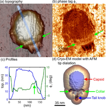
| Researchers in the United States and Spain have discovered that an atomic force microscope - a tool widely used in nanoscale imaging - works differently in watery environments, a step toward better using the instrument to study biological molecules and structures. The researchers demonstrated their new understanding of how the instrument works in water to show details of the mechanical properties of a virus called Phi29. The images in "a" and "c" show the topography, and the image in "b" shows the different stiffness properties of the balloonlike head, stiff collar and hollow tail of the Phi29 virus, called a bacteriophage because it infects bacteria. (C. Carrasco-Pulido, P. J. de Pablo, J. Gomez-Herrero, Universidad Autonoma de Madrid, Spain) |
Abstract:
Origins of Phase Contrast in the Atomic Force Microscope in Liquids
John Melchera, Carolina Carrascob,c, Xin Xua, José L. Carrascosad, Julio Gómez-Herrerob,
Pedro José de Pablob, and Arvind Ramana
(a) School of Mechanical Engineering and Birck Nanotechnology Center, Purdue University, West Lafayette, IN 47907; (b) Departamento de Física de la Materia Condensada C-III, Universidad Autónoma de Madrid, 28049 Madrid, Spain; (c) Instituto de Ciencia de Materiales de Madrid, Centro National de Biotecnología, Consejo Superior de Investigaciones Científicas, 28049 Madrid, Spain; (d) Departmento de Estructura de Macomoléculas, Centro National de Biotecnología, Consejo Superior de Investigaciones Científicas, 28049 Madrid, Spain
We study the physical origins of phase contrast in dynamic atomic force microscopy (dAFM) in liquids where low-stiffness microcantilever probes are often used for nanoscale imaging of soft biological samples with gentle forces. Under these conditions, we show that the phase contrast derives primarily from a unique energy flow channel that opens up in liquids due to the momentary excitation of higher eigenmodes. Contrary to the common assumption, phase-contrast images in liquids using soft microcantilevers are often maps of short-range conservative interactions, such as local elastic response, rather than tip-sample dissipation. The theory is used to demonstrate variations in local elasticity of purple membrane and bacteriophage _29 virions in buffer solutions using the phase-contrast images.
Discovery to aid study of biological structures, molecules
WEST LAFAYETTE, IN | Posted on August 11th, 2009Researchers in the United States and Spain have discovered that a tool widely used in nanoscale imaging works differently in watery environments, a step toward better using the instrument to study biological molecules and structures.
The researchers demonstrated their new understanding of how the instrument - the atomic force microscope - works in water to show detailed properties of a bacterial membrane and a virus called Phi29, said Arvind Raman, a Purdue professor of mechanical engineering.
"People using this kind of instrument to study biological structures need to know how it works in the natural watery environments of molecules and how to interpret images," he said.
An atomic force microscope uses a tiny vibrating probe to yield information about materials and surfaces on the scale of nanometers, or billionths of a meter. Because the instrument enables scientists to "see" objects far smaller than possible using light microscopes, it could be ideal for studying molecules, cell membranes and other biological structures.
The best way to study such structures is in their wet, natural environments. However, the researchers have now discovered that in some respects the vibrating probe's tip behaves the opposite in water as it does in air, said Purdue mechanical engineering doctoral student John Melcher.
Purdue researchers collaborated with scientists at three institutions in Madrid, Spain: Universidad Autónoma de Madrid, Instituto de Ciencia de Materiales de Madrid and the Centro Nacional de Biotecnología.
Findings, which were detailed in a paper appearing online last week in the U.S. publication Proceedings of the National Academy of Sciences, are related to the subtle differences in how the instrument's probe vibrates. The probe is caused to oscillate by a vibrating source at its base. However, the tip of the probe oscillates slightly out of synch with the oscillations at the base. This difference in oscillation is referred to as a "phase contrast," and the tip is said to be out of phase with the base.
Although these differences in phase contrast reveal information about the composition of the material being studied, data can't be properly interpreted unless researchers understand precisely how the phase changes in water as well as in air, Raman said.
If the instrument is operating in air, the tip's phase lags slightly when interacting with a viscous material and advances slightly when scanning over a hard surface. Now researchers have learned the tip operates in the opposite manner when used in water: it lags while passing over a hard object and advances when scanning the gelatinous surface of a biological membrane.
Researchers deposited the membrane and viruses on a sheet of mica. Tests showed the differing properties of the inner and outer sides of the membrane and details about the latticelike protein structure of the membrane. Findings also showed the different properties of the balloonlike head, stiff collar and hollow tail of the Phi29 virus, called a bacteriophage because it infects bacteria.
"The findings suggest that phase contrast in liquids can be used to reveal rapidly the intrinsic variations in local stiffness with molecular resolution, for example, by showing that the head and the collar of an individual virus particle have different stiffness," Raman said.
The research was funded by the National Science Foundation and was conducted at the Birck Nanotechnology Center in Purdue's Discovery Park. The biological membrane images were taken at Purdue, and the virus studies were performed at the Universidad Autónoma de Madrid.
The paper was authored by Melcher; Carolina Carrasco, a postdoctoral researcher at Universidad Autónoma de Madrid and the Instituto de Ciencia de Materiales de Madrid; Purdue postdoctoral researcher Xin Xu; José L. Carrasco, a researcher at Departmento de Estructura de Macomoléculas, Centro Nacional de Biotecnología, Consejo Superior de Investigaciones Científicas; Julio Gómez-Herrero and Pedro José de Pablo, both researchers from Universidad Autónoma de Madrid; and Raman.
####
For more information, please click here
Contacts:
Writer: Emil Venere
(765) 494-4709
Sources: Arvind Raman
(765) 494-5733
John Melcher
Purdue News Service
(765) 494-2096
Copyright © Purdue University
If you have a comment, please Contact us.Issuers of news releases, not 7th Wave, Inc. or Nanotechnology Now, are solely responsible for the accuracy of the content.
| Related News Press |
News and information
![]() Decoding hydrogen‑bond network of electrolyte for cryogenic durable aqueous zinc‑ion batteries January 30th, 2026
Decoding hydrogen‑bond network of electrolyte for cryogenic durable aqueous zinc‑ion batteries January 30th, 2026
![]() COF scaffold membrane with gate‑lane nanostructure for efficient Li+/Mg2+ separation January 30th, 2026
COF scaffold membrane with gate‑lane nanostructure for efficient Li+/Mg2+ separation January 30th, 2026
![]() MXene nanomaterials enter a new dimension Multilayer nanomaterial: MXene flakes created at Drexel University show new promise as 1D scrolls January 30th, 2026
MXene nanomaterials enter a new dimension Multilayer nanomaterial: MXene flakes created at Drexel University show new promise as 1D scrolls January 30th, 2026
Imaging
![]() ICFO researchers overcome long-standing bottleneck in single photon detection with twisted 2D materials August 8th, 2025
ICFO researchers overcome long-standing bottleneck in single photon detection with twisted 2D materials August 8th, 2025
Nanomedicine
![]() New molecular technology targets tumors and simultaneously silences two ‘undruggable’ cancer genes August 8th, 2025
New molecular technology targets tumors and simultaneously silences two ‘undruggable’ cancer genes August 8th, 2025
![]() New imaging approach transforms study of bacterial biofilms August 8th, 2025
New imaging approach transforms study of bacterial biofilms August 8th, 2025
![]() Cambridge chemists discover simple way to build bigger molecules – one carbon at a time June 6th, 2025
Cambridge chemists discover simple way to build bigger molecules – one carbon at a time June 6th, 2025
![]() Electrifying results shed light on graphene foam as a potential material for lab grown cartilage June 6th, 2025
Electrifying results shed light on graphene foam as a potential material for lab grown cartilage June 6th, 2025
Discoveries
![]() From sensors to smart systems: the rise of AI-driven photonic noses January 30th, 2026
From sensors to smart systems: the rise of AI-driven photonic noses January 30th, 2026
![]() Decoding hydrogen‑bond network of electrolyte for cryogenic durable aqueous zinc‑ion batteries January 30th, 2026
Decoding hydrogen‑bond network of electrolyte for cryogenic durable aqueous zinc‑ion batteries January 30th, 2026
![]() COF scaffold membrane with gate‑lane nanostructure for efficient Li+/Mg2+ separation January 30th, 2026
COF scaffold membrane with gate‑lane nanostructure for efficient Li+/Mg2+ separation January 30th, 2026
Announcements
![]() Decoding hydrogen‑bond network of electrolyte for cryogenic durable aqueous zinc‑ion batteries January 30th, 2026
Decoding hydrogen‑bond network of electrolyte for cryogenic durable aqueous zinc‑ion batteries January 30th, 2026
![]() COF scaffold membrane with gate‑lane nanostructure for efficient Li+/Mg2+ separation January 30th, 2026
COF scaffold membrane with gate‑lane nanostructure for efficient Li+/Mg2+ separation January 30th, 2026
Tools
![]() Metasurfaces smooth light to boost magnetic sensing precision January 30th, 2026
Metasurfaces smooth light to boost magnetic sensing precision January 30th, 2026
![]() From sensors to smart systems: the rise of AI-driven photonic noses January 30th, 2026
From sensors to smart systems: the rise of AI-driven photonic noses January 30th, 2026
![]() Japan launches fully domestically produced quantum computer: Expo visitors to experience quantum computing firsthand August 8th, 2025
Japan launches fully domestically produced quantum computer: Expo visitors to experience quantum computing firsthand August 8th, 2025
Research partnerships
![]() Lab to industry: InSe wafer-scale breakthrough for future electronics August 8th, 2025
Lab to industry: InSe wafer-scale breakthrough for future electronics August 8th, 2025
![]() HKU physicists uncover hidden order in the quantum world through deconfined quantum critical points April 25th, 2025
HKU physicists uncover hidden order in the quantum world through deconfined quantum critical points April 25th, 2025
|
|
||
|
|
||
| The latest news from around the world, FREE | ||
|
|
||
|
|
||
| Premium Products | ||
|
|
||
|
Only the news you want to read!
Learn More |
||
|
|
||
|
Full-service, expert consulting
Learn More |
||
|
|
||


